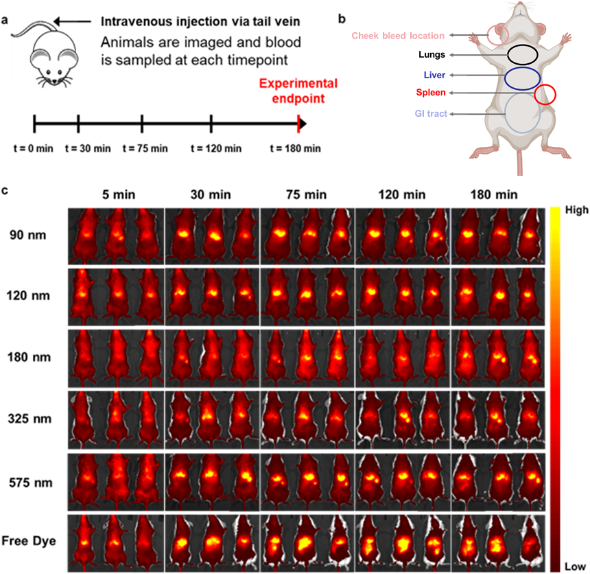
Figure 7. In vivo biodistribution of Cy7-labeled nanoparticles and a free dye control in BALB/c mice.

(a). Dosing, blood sampling, and imaging schedule. (b) Overview of organ locations in mouse. Adapted from Biorender42 (c). Live imaging of Cy7-labeled hemostatic nanoparticles and free dye control over three hours. Sizes are expressed as the average of each size range for space considerations. Dark red/black denote lower levels of fluorescence, while yellow indicates accumulation in a particular area.
