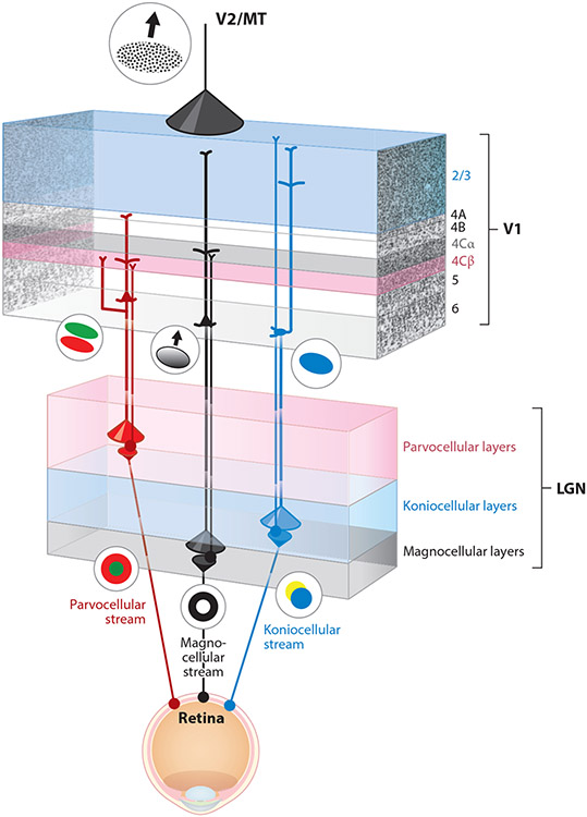
Figure 1.
Feedforward and feedback parallel pathways in the early visual system of primates. Retinal ganglion cells conveying visual information to the dorsal lateral geniculate nucleus (LGN) of the thalamus are separated into three major parallel processing streams: the parvocellular (red), magnocellular (black), and koniocellular (blue) streams. These retinal ganglion cells (filled colored circles at bottom) project axons to separated parvocellular (pink), magnocellular (gray), and intercalated koniocellular (blue) layers of the LGN. LGN relay neurons (filled colored circles in LGN) display distinctive receptive field properties driven by their retinal inputs: red–green center–surround organization among parvocellular LGN neurons, On–Off (black–white) center–surround organization among magnocellular LGN neurons, and blue–yellow opponent organization among koniocellular neurons. The geniculocortical axons of LGN relay neurons target segregated layers within the primary visual cortex (V1): Parvocellular geniculocortical axons (red thin line) mainly target layer 4Cβ (pink); magnocellular geniculocortical axons (black thin line) mainly target layer 4Cα (gray); and koniocellular geniculocortical axons (blue thin line) target layer 1, the blobs in layer 2/3, and layer 4A. Geniculocortical recipient neurons in these V1 layers also display distinctive receptive field properties driven by their geniculocortical inputs: Parvocellular-recipient V1 neurons are simple cells with separable red–green receptive field subregions, magnocellular-recipient V1 neurons are simple and complex cells with orientation tuning and direction selectivity, and koniocellular-recipient V1 neurons are responsive to blue color. Corticogeniculate neurons located in layer 6 of V1 provide feedback from V1 to the LGN. Corticogeniculate neurons are also separated into the same parallel streams, and their receptive field properties reflect their feedforward stream-specific inputs. There are parvocellular-projecting corticogeniculate neurons (red pyramidal cell in upper layer 6 of V1), magnocellular-projecting corticogeniculate neurons (black pyramidal cell in lower layer 6), and koniocellular-projecting corticogeniculate neurons (blue tilted cell at the bottom of layer 6). The distributions of corticogeniculate axon terminals in the LGN (red, black, and blue downward cones) are broader compared to the distributions of retinogeniculate axon terminals in the LGN (red, black, blue upward cones). Similarly, the distribution of terminals from corticocortical feedback axons (top black cone) are also more broadly distributed relative to feedforward and local axon terminal distributions within V1. Corticocortical feedback originates in extrastriate visual areas such as the secondary visual cortex (V2) and the middle temporal area (MT), in which neurons are responsive to more complex visual features including texture and stimulus motion.

