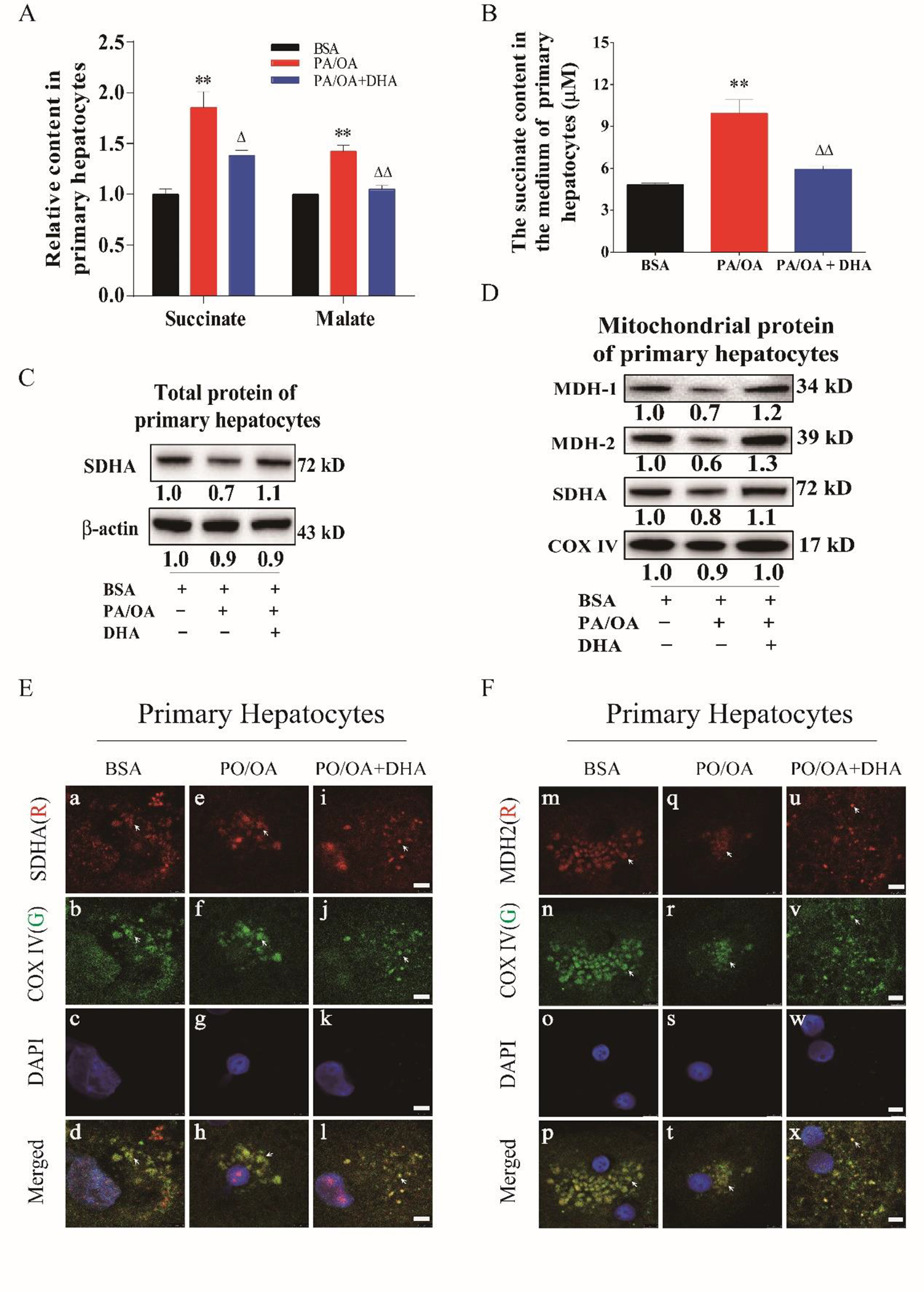
Fig. 3. Down-regulation of MDH2 and SDHA expression in primary hepatocytes exposed to fatty acid overload.

A. Succinate and malate content in primary hepatocytes. B. Succinate content in culture medium of primary hepatocytes exposed to PA/OA overload plus/minus DHA supplementation. C. Protein levels of SDHA in in primary hepatocytes in response to fatty acid overload. D. Protein levels of MDH1/2 in primary hepatocytes in response to fatty acid overload. E. Immunocytochemical staining of SDHA in primary hepatocytes in exposure to fatty acid overload. F. Immunocytochemical staining of MDH2 in primary hepatocytes in exposure to fatty acid overload. Primary hepatocytes were exposed to a combination overload of palmitic acid (PA) with oleic acid (OA). PA and OA was first dissolved in methanol and added to medium at the final concentration of 200μM and 400μM. DHA was first dissolved in ethanol and added to medium at the final concentration of 400 μM. DHA was added to medium 15 minutes before PA/OA. The experiment was repeated at least 3 times. All data were expressed as mean ± SEM. ** p < 0.01 compared to BSA. △, △△ p < 0.05 and 0.01 compared to PA/OA. The ANOVA variance test was used to compare between groups, and the LSD test was used for multiple comparisons between two given groups.
