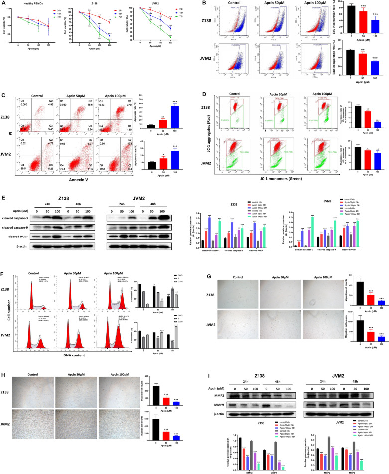
Fig. 3.
CDC20 inhibitor apcin could inhibit cell proliferation, migration and invasion, and induce cell apoptosis and cell cycle arrest in Z138 and JVM2 cells. A After healthy PBMCs, Z138 and JVM2 cells exposed to 50 μM, 100 μM and 200 μM apcin for 24 h, 48 h and 72 h, cell viability was assessed by CCK-8 assay. Cell viability at each time point was defined as the percentage obtained by dividing the OD value of the treatement group by the OD value of the corresponding untreated group at 450 nm. B After Z138 and JVM2 cells treated with 50 μM and 100 μM apcin for 48 h, EdU incorporation rate was detected by flow cytometry to determine the cell proliferation condition. C The apoptosis rate was measured by flow cytometry based on Annexin V-FITC/PI staining after Z138 and JVM2 cells treated with 50 μM and 100 μM apcin for 48 h. The total apoptosis rate was the sum of the early apoptosis rate (right lower quadrant) and the late apoptosis rate (right upper quadrant). D The changes of MMP was estimated by flow cytometry based on JC-1 fluorescent probe after Z138 and JVM2 cells incubated with 50 μM and 100 μM apcin for 48 h. The results were presented as the ratio of mean red fluorescence intensity to mean green fluorescence intensity. Decreased ratio indicated the decrease in MMP, which was also a hallmark event of early apoptosis. E Expression of Apoptosis-related proteins by WB analysis after 24 h and 48 h apcin treatment in Z138 and JVM2 cells. F The proportion of G0/G1, S and G2/M phases in the cell cycle was analyzed by PI flow cytometry after Z138 and JVM2 cells treated with 50 μM and 100μmM apcin for 48 h (Z138) or 72 h (JVM2). G, H The effect of apcin on cell migration (G) and invasion (H) was confirmed by Transwell assays after Z138 and JVM2 cells exposed to 50 μM and 100 μM apcin for 48 h. Images were captured by an inverted microscope (× 10 magnification). (I) Migration and invasion-related proteins was analyzed by WB after 24 h and 48 h apcin treatment in Z138 and JVM2 cells. The above data were obtained from at least three independent experiments and presented as mean ± SD. *P < 0.05, **P < 0.01, ***P < 0.001 compared with the control group

