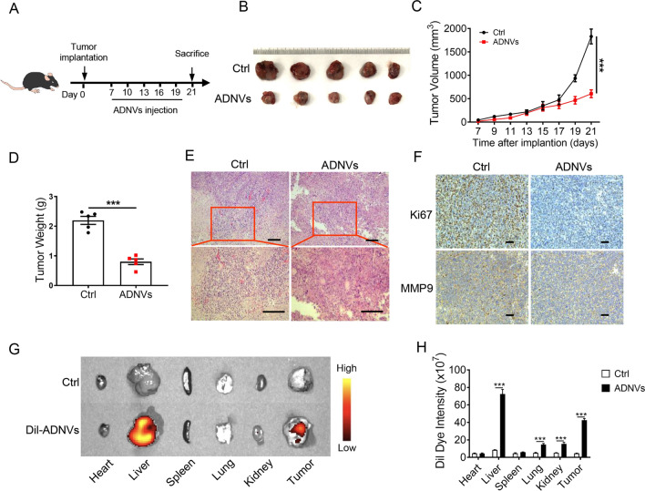
Fig. 2.
ADNVs inhibit lung cancer growth in mice. A The simplified experimental scheme. C57BL/6 mice (n = 5) were implanted with LLC cells for 7 d, and then treated with ADNVs (25 mg/kg, i.p.) once every 3 d for a successive 2 week. Mice were sacrificed and tumors were collected at day 21. B Gross photos of tumors at the end of experiments. C Tumor growth profiles in tumor-bearing mice treated PBS or ADNVs. ***p < 0.001 (Two-way ANOVA and Bonferroni post-tests). D Tumor weights in mice treated with either PBS or ADNVs. ***p < 0.001 (Student’s t-test). E H & E staining of tumor tissues (Scale bar = 100 μm). F Ki67 and MMP9 staining of tumor tissues (Scale bar = 100 μm). (G, H) ADNVs were stained with Dil and injected into tumor-bearing mice (25 mg/kg, i.p.). Biodistribution of ADNVs was determined by scanning mice (G), and the quantitative analysis (H). ***p < 0.001 (One-way ANOVA and Tukey’s significant difference post hoc test). The results are representative data from one of three independent experiments. Shown are representative images, and the data are presented as means ± SEM

