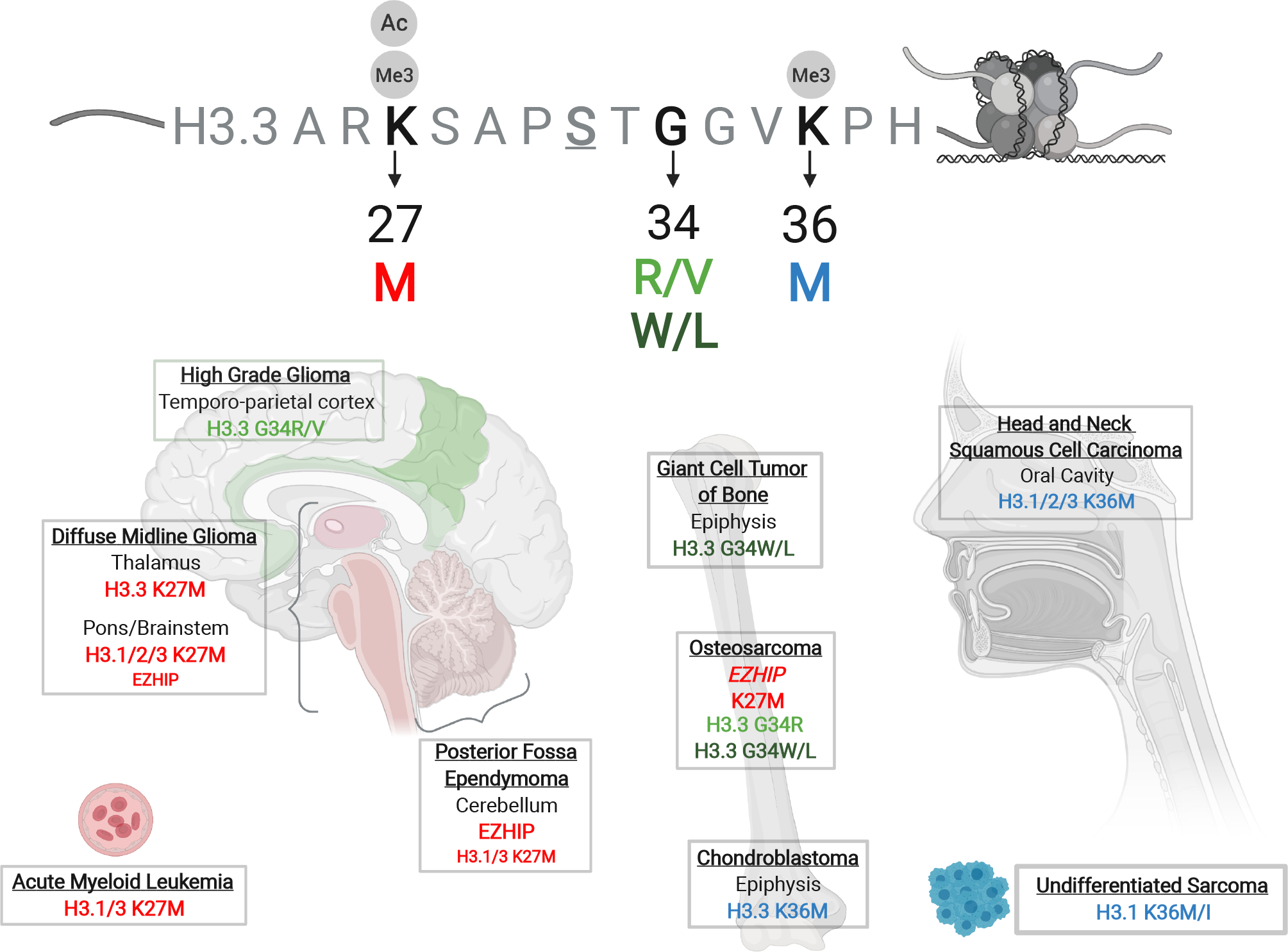
Figure 1. Histone mutations in cancers.

Schematic of the histone H3.3 tail above, highlighting key residues (K27, G34, K36) recurrently mutated in cancers and their associated post-translational modifications. Depicted below is the regional tissue specificity of oncohistone mutations and their occurrence in specific cancer types.
