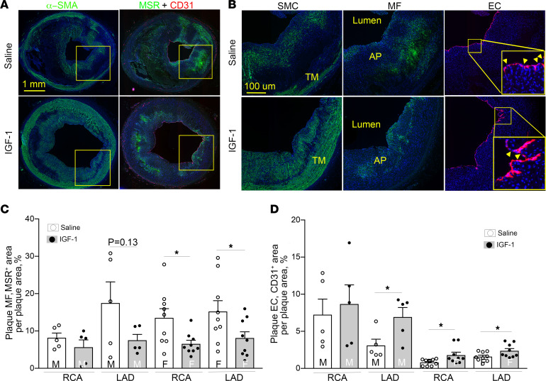
Figure 4. IGF-1 suppresses MF-like cells and upregulates EC-like cells in coronary plaques.
Serial RCA and LAD sections were immunostained with α–smooth muscle actin (α-SMA), macrophage scavenger receptor A (MSR), and CD31 antibody to identify SMC-like, MF-like, and EC-like cells, respectively. The primary antibody signal was amplified by biotin/streptavidin or tyramide systems conjugated to Alexa Fluor 488 (for α-SMA and MSR) or Alexa Fluor 594 (CD31). (A) Representative images of RCA sections obtained from IGF-1– or saline-injected FH females. Yellow square outlines plaque area magnified in B. (B) SMC, MF, and EC marker–immunopositive cells. Yellow arrows in insert indicate breaks in endothelial layer. (C and D) Quantitative data. n = 5 per RCA or LAD per group for males and n = 9 for females. *P < 0.05 vs. saline based on unpaired 2-tailed t test.

