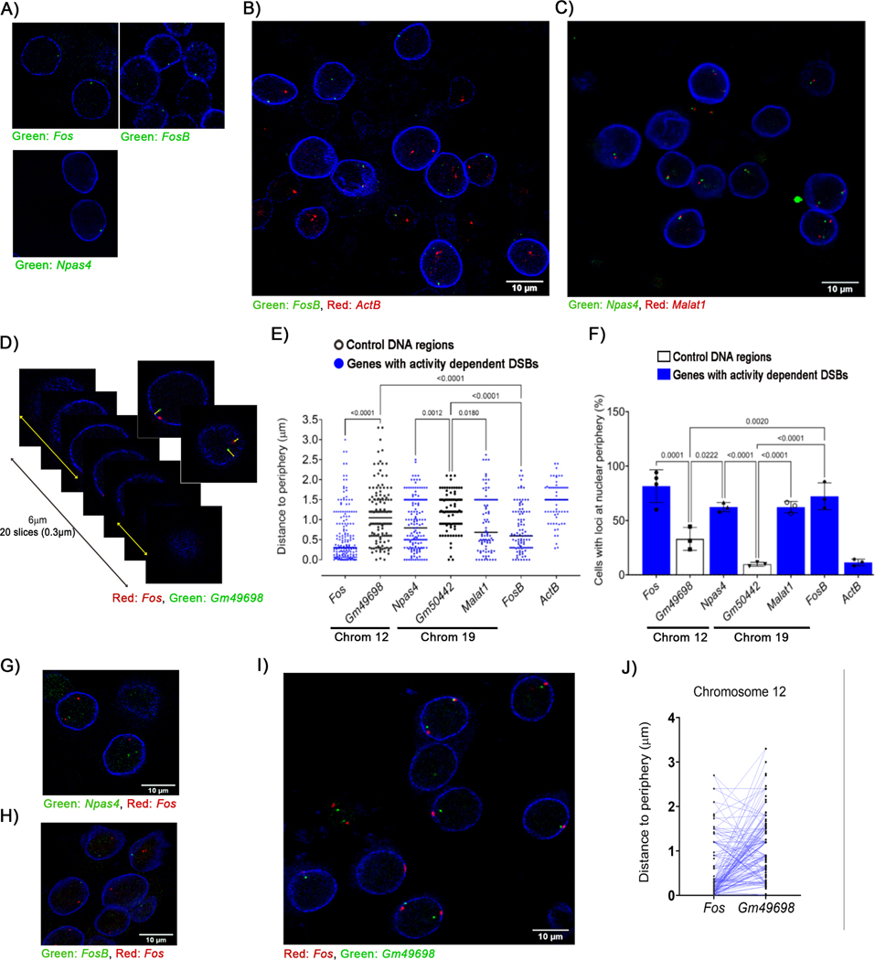
Figure 6. Sites that incur activity-induced DSBs preferentially localize to the nuclear periphery in neurons.
DNA immuno-FISH was performed using fluorescently labeled BAC probes that hybridize with genomic regions containing the indicated genes and antibodies against lamin A/C. Probes were used to assess visualize either a single locus (A) or two distinct loci simultaneously as indicated (B, C, D, G, H, and I) Images show representative optical slices from z-stacks. (D – F) (D) Distance of the indicated loci to the nuclear rim was measured as in Figure S6A). (E) Quantification of the distance of each probe from the nuclear periphery as shown in (D). (F) Loci that were located at a distance < 0.5 μm from lamin A/C were defined as being localized to the nuclear periphery and the percentage of cells with at least one copy of the probed region located at the nuclear periphery were plotted. (J) The shortest distance to the periphery (either XY or Z) for Fos and Gm49698, which are both located on chromosome 12, were compared on a single chromosome level and plotted (linked by lines).
See also Figure S6.

