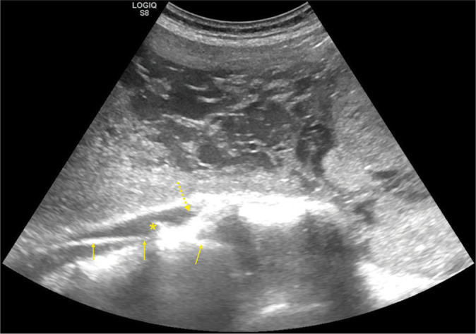
Figure 1:

Coronal oblique plane ultrasound image obtained from the left anterolateral aspect with compression to displace the uterus to the right side with the probe after placing an angiographic metallic guidewire via right femoral access showing the guidewire as a linear intravascular reflective structure (yellow arrows) in the infrarenal aorta and right common iliac artery, the location of the aortic bifurcation (asterisk), and the proximal segment of the left common iliac artery (dashed arrows).
