
Figure 1.
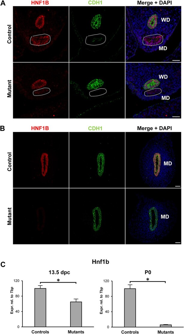
Hnf1b ablation in Mullerian duct epithelium. (A) In control animals, HNF1B was expressed in both the Wolffian ducts (WD) and the Müllerian ducts (MD, dashed line) at 13.5 dpc. In mutants, HNF1B expression was lost specifically in the MD. (B) At P0, only the MD remains in female animals, and immunohistochemistry showed no expression in the mutants. The epithelial marker CDH1/E-cadherin was used to visualize the WD at 13.5dpc and the MD at P0. Scale bar = 25 μm. (C) Real-time PCR of whole mesonephric tissues showed a reduction in Hnf1b expression at 13.5 dpc in mutant samples, consistent with the presence of the Hnf1b-expressing WD. At P0, expression of Hnf1b was lost in the mutants. * = P < 0.05.