Figure 3.
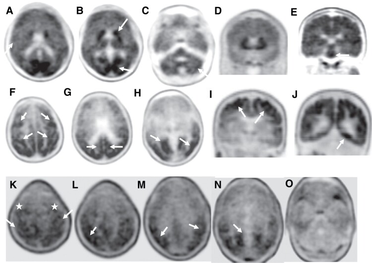
Amyloid and tau PET images in affected family members. (1. Top row) Florbetapir PET images of Case BII in axial (A–C) and coronal (D and E) planes showing loss of grey–white matter differentiation (A, arrow) with multiple areas of high cortical uptake particularly in the posterior cingulate gyri, parietal and occipital lobes with very high uptake in the basal ganglia and brainstem (B and E, arrows). Patchy uptake is also seen in the grey matter of the cerebellum (C, arrow). (2. Middle row) Flortaucipir PET images of case BI; axial (F–H) and coronal (I and J) planes show extensive tau uptake in the neocortical regions. There are bilateral frontal and parietal uptakes including the peri-Rolandic regions (F and I, arrows), posterior cingulate gyri (G, arrow) and premotor cortices (F, top arrows). There is also extensive binding in the lateral and medial occipital areas bilaterally (G and J, arrows). (3. Bottom row) Flortaucipir PET-CT images of case BII; axial images showing patchy uptake in neocortical regions, including parietal, occipital, posterolateral temporal lobes (L–O, arrows) and also in the peri-Rolandic regions (K, arrows) and premotor cortices (K, white stars). No cerebellar uptake identified.

