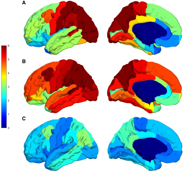
Figure 4.
SUVRs for flortaucipir PET in persons affected by autosomal dominant Alzheimer’s disease mutations. The averaged SUVR for both cerebral hemispheres shown on the left hemisphere of the fsaverage template from all subjects in each group. For each subject, SUVR value was first averaged for each region of interest and then left and right hemispheres were averaged shown here on the fsaverage template for each group. The top row (A) is data from the two brothers affected by the F388S mutation, the middle row (B) is derived from seven persons affected by the A431E mutation in PSEN1, and the bottom row (C) is derived from three persons affected by autosomal dominant Alzheimer’s disease mutations that do not cause spastic paraparesis. Note the variable involvement of the Rolandic cortex associated with presence or absence of SP but consistent involvement of the precuneus across groups.

