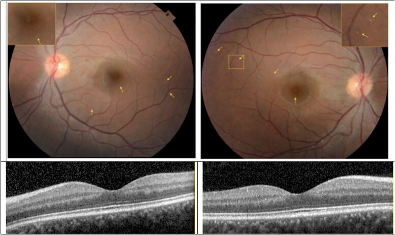
Figure 5.
Colour fundus photograph and optical coherence tomography (OCT) of the left (left panels) and right eyes (right panels) of subject BII. The optic disc appears with sharp margins and normal colour in both eyes. The retinal vessels are largely unremarkable except for possibly mild tortuosity and focal areas of attenuation of the large calibre retinal arteries in each eye. The fundus pigmentation is notable for small (∼25 microns), focal lesions in each eye some of which are denoted by the small arrows. Inset panels in the fundus photographs demonstrate high magnification of a region with the lesions. OCT sections through the fovea of each are illustrated. There was no evidence of retinal pigment epithelium or intraretinal pathology on any OCT section that corresponded with lesions on fundus photography.

