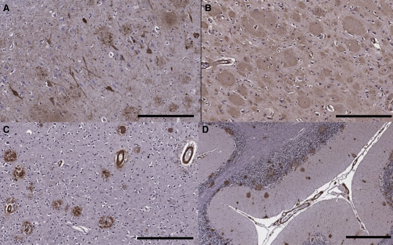
Figure 6.
Immunohistochemistry on neuropathological specimens from the patient with the F388S PSEN1 mutation. (A) The hippocampus shows abundant neurofibrillary tangles and neuritic plaques on tau (AT8) immunohistochemistry (scale bar = 300 μm). (B) Florid amyloid plaques with ‘cotton wool’ morphology in the frontal cortex on Aβ42 immunostain (300 μm). (D) Immunohistochemistry for Aβ40 highlights severe CAA in the occipital cortex as well as scattered plaques with dense cores (300 μm). (D) Many amyloid plaques as well as CAA seen in the cerebellar cortex on Aβ42 immunostain (scale bar = 600 μm).

