Fig. 9.
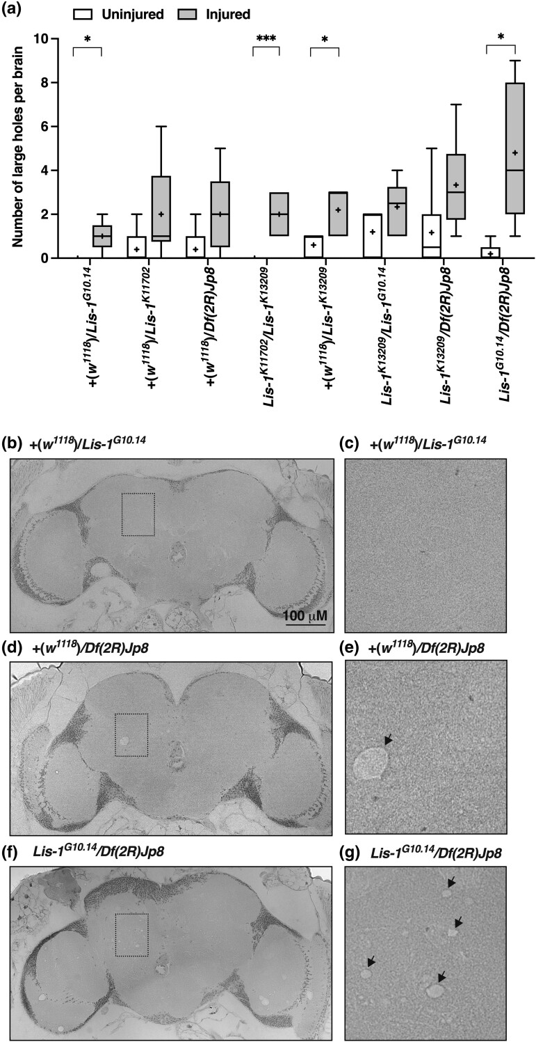
Lis-1 mutations enhance neurodegeneration in the brain following TBI. a) The number of large (>5 μM) holes in the brain of female Lis-1 mutants of the indicated genotypes at two weeks after injury at 0–7 days old (injured) or at the same age in the absence of injury (uninjured) (n = 4). Lis-1 heterozygotes were generated by crossing Lis-1 mutants to the w1118 line. Brain sections chosen for analysis were at equivalent depths in the brain. Genotypes are ordered from low to high, based on the mean number of holes per brain in injured flies. Symbols indicate the following: box, second and third quartiles of data; +, mean; horizonal bar, median; and whiskers, minimum and maximum data points. (b–d) Representative images of sections of fly brains from flies of the indicated genotypes and injury status. (e–g) High-magnification images of boxed regions in b–d, respectively. Arrows indicate large holes.

