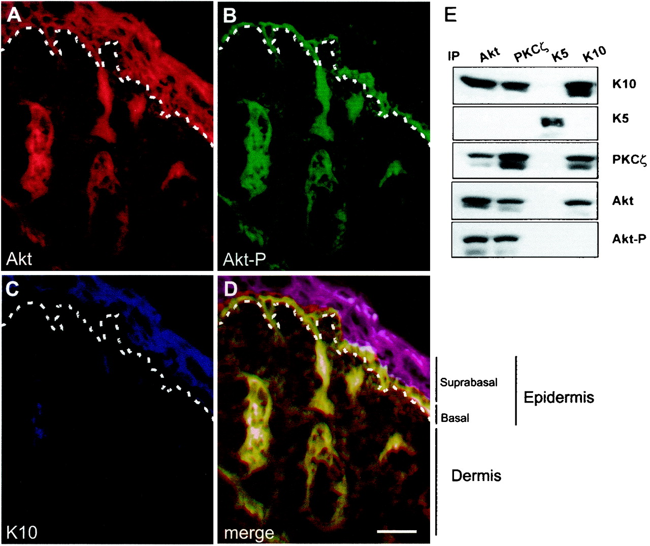
FIG. 6.

Distribution of Akt, Akt-P, and K10 in newborn mouse skin. (A to D) Frozen sections of newborn mouse skin were processed for immunofluorescence using antibodies against Akt (A), Akt-P (B), and K10 (C). The three immunofluorescence channels (Texas red, fluorescein isothiocyanate, and AMCA, respectively) are shown merged in panel D. Note that Akt is present throughout all the epidermal layers, whereas phosphorylated Akt is present exclusively in the basal layer and K10 is restricted to differentiated suprabasal layers. This shows that Akt and K10 are coexpressed in suprabasal cells but that only basal, K10-negative cells have active, phosphorylated Akt. Dashed lines, epidermal-dermal boundary. (E) Immunoprecipitation-Western blot experiments to biochemically confirm the K10-Akt-PKCζ interaction in skin. Whole-skin extracts were immunoprecipitated (IP) with the indicated antibodies and probed by Western blotting with the antibodies to proteins indicated at the right. Note the presence of K10, but not K5, in Akt and PKCζ immunoprecipitates and vice versa, whereas phosphorylated Akt is absent from K10 immunoprecipitates. Also note the absence of the kinases in K5 immunoprecipitates. Bar, 100 μm.
