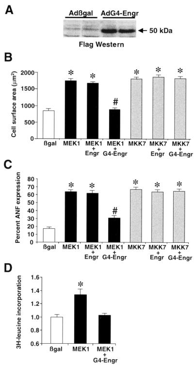
FIG. 7.
GATA4-engrailed blocks MEK1-ERK1/2-induced cardiomyocyte hypertrophy. (A) Western blotting with Flag monoclonal antibody demonstrates a protein of the predicted size from AdG4-Engr-infected cardiomyocytes. (B) Cell surface areas were quantified from each of the indicated adenoviral infected cardiomyocyte cultures. *, P < 0.05 versus Adβgal; #, P < 0.05 versus AdMEK1 plus AdEngr. (C) The percentage of cells expressing ANF protein was quantified from each of the indicated adenoviral infected cardiomyocyte cultures. *, P < 0.05 versus Adβgal; #, P < 0.05 versus AdMEK1 plus AdEngr. (D) [3H]leucine incorporation was measured in Adβgal-, AdMEK1-, and AdMEK1-plus-AdG4-Engr-infected cardiomyocytes. *, P < 0.05 versus Adβgal. All data represent the averages of three independent experiments.

