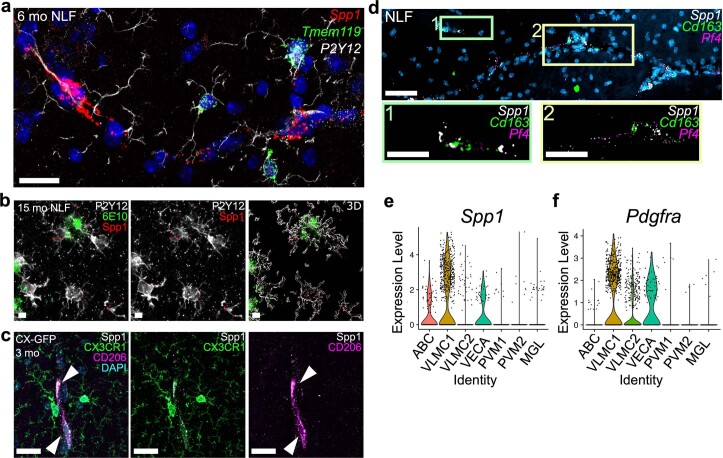
Extended Data Fig. 2. Spp1 is expressed in perivascular cells.
(a) Representative confocal image showing Spp1 and Tmem119 mRNA expression within P2Y12+ microglia of 6 mo AppNL-F SLM as characterized by smFISH-IHC. Scale bar represents 20 µm. Data representative of n = 8 AppNL-F mice examined over 3 independent experiments. (b) Representative confocal image showing Spp1 mRNA in P2Y12 microglia associated with 6E10+ plaques in 15 mo AppNL-F SLM. Scale bar represents 5 µm. Data representative of n = 3 AppNL-F mice examined over 2 independent experiments. (c) Representative confocal images of SPP1 protein expression in CD206+ PVMs (arrow head) and CX3CR1GFP myeloid cells of 3 mo Cx3cr1GFP/WT SLM86. Scale bar represents 20 µm. Data are representative of n = 2 Cx3cr1GFP/WT mice examined over at least 3 independent experiments. (d) Representative confocal image showing Spp1 mRNA expression occasionally colocalizing with pan-PVM markers Cd163 and Pf4 in 6 mo AppNL-F SLM as identified by smFISH. Scale bar represents 50 µm. Data representative of n = 6 AppNL-F mice examined over 2 independent experiments. (e, f) Violin plots of Spp1 (e) and Pdgfra (f) expression reanalyzed from Zeisel et al.37. VLMC, vascular leptomeningeal cells; ABC, Arachnoid barrier cells; VECA, Arterial vascular endothelial cells; PVM, Perivascular macrophage; MGL, Microglia.

