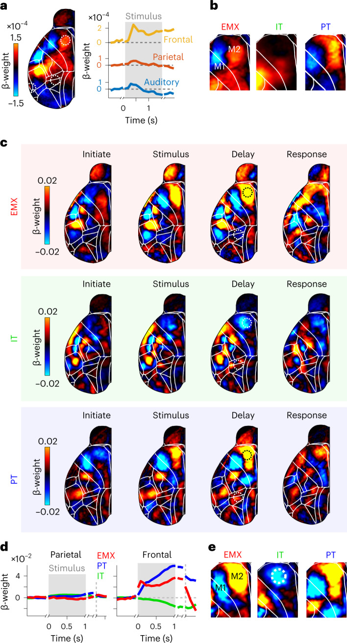
Fig. 6. The temporal dynamics of choice-related activity differ across pyramidal neuron types.

a, Left, averaged contralateral choice kernels for EMX mice during the delay period. Positive weights indicate increased choice-related activity for contralateral choices, while negative weights indicate decreased choice-related activity. Right, choice-related activity in auditory (blue), parietal (red) and frontal cortices (yellow). Traces are realigned to the initiation, stimulus, delay and response periods, indicated by gaps in weight traces. b, Zoomed-in map for delay-period frontal choice kernels of EMX, IT and PT neurons. c, Cortical maps of contralateral choice weights for different trial episodes. Several areas in anterior cortex showed clear choice signals. d, Baseline-corrected decoder weights in parietal (left) and frontal (right) cortices throughout the trial. Conventions as in a. Dashed circles in the delay maps of c show the parietal and frontal locations used to compute the traces. e, Zoomed-in map for frontal delay-period decoder weights of EMX, IT and PT mice. Dashed circle shows the ALM.
