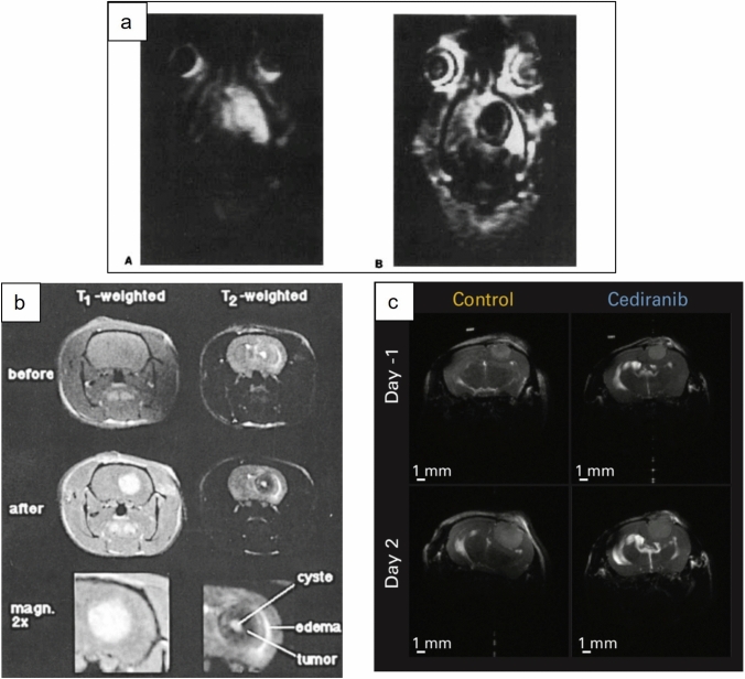
Fig. 1.
Representative MR images of different edema patterns obtained in animal models. a an example of infiltrative tumor edema captured on T1 IR snapshot FLASH imaging from rats bearing F98 glioma, reproduced with permission[52]. b an example of a peritumoral ‘halo’ of edema, demonstrated in T1- and T2-weighted MR images in Fischer rats bearing F98 glioma, reproduced with permission[30]. c an example of tumor with no detectable edema on T2-weighted MR in nude mice bearing U87 tumors, reproduced with permission [25]

