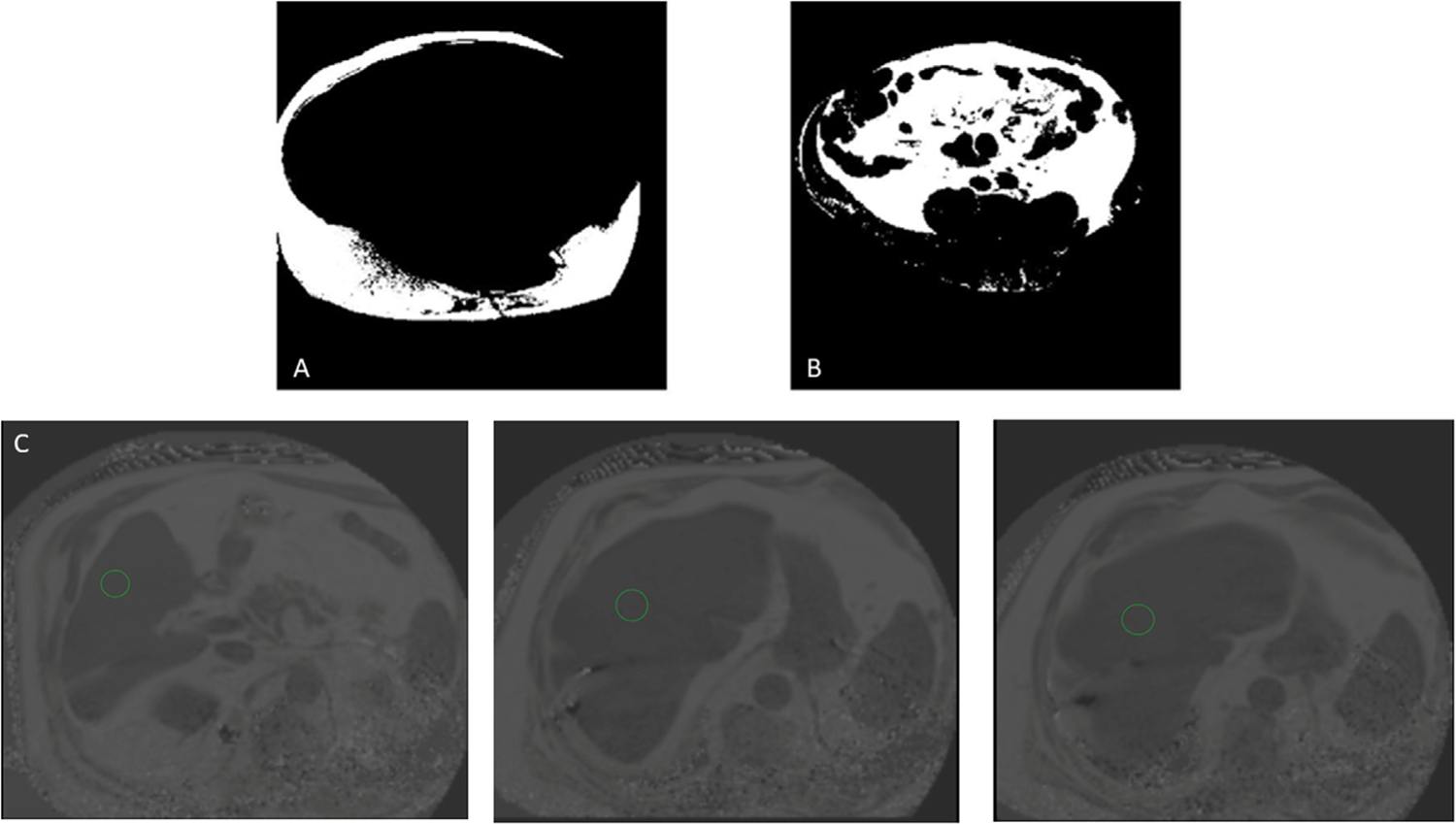
Fig. 2.

Magnetic resonance imaging of abdominal and liver adiposity in a participant with HFpEF.
Abdominal fat was sectioned into its subcutaneous (A) and visceral (B) components using a semi-automated MATLAB tool. (C) Liver fat fraction was estimated by averaging the magnetic resonance imaging proton-density fat fraction across three axial slices for the region of interest circled in green. HFpEF = heart failure with preserved ejection fraction.
