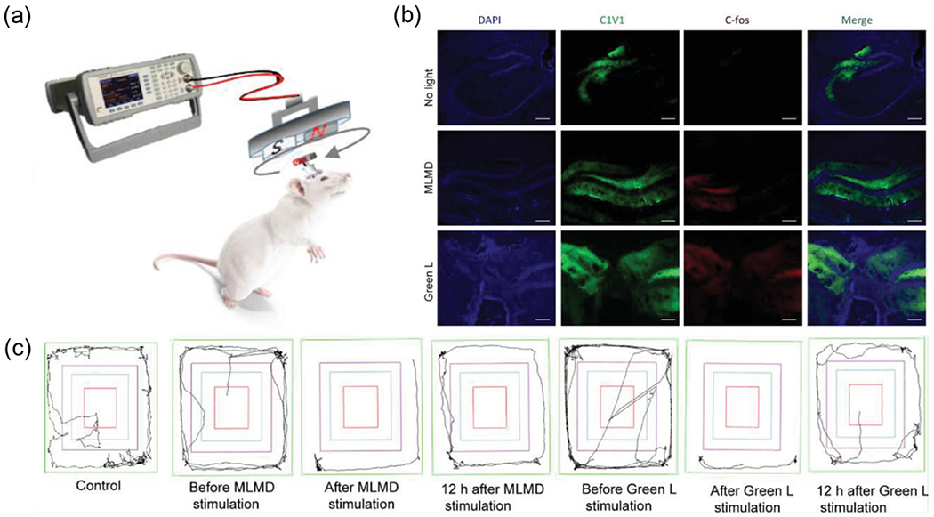
Fig.8. Wireless optogenetics enabled by a MLMD.

(a) Schematic showing MLMD device implanted in the primary motor cortex (M1) and the wireless neuromodulation by MLMD in free-moving mice. (b) The confocal images of C1V1 and C-fos staining in hippocampus regions of the mouse brain in different groups. All scale bars represent 200 μm. (c) The displacement trajectories of mice with different neuromodulation conditions. Reproduced from [88] with permission from Wiley-VCH, Copyright 2021.
