
Figure 1. Exome and targeted sequencing in nephrolithiasis (NL)/nephrocalcinosis (NC) subjects revealed dominant OXGR1 variants, which impair Ca2+ uptake in response to ligand.
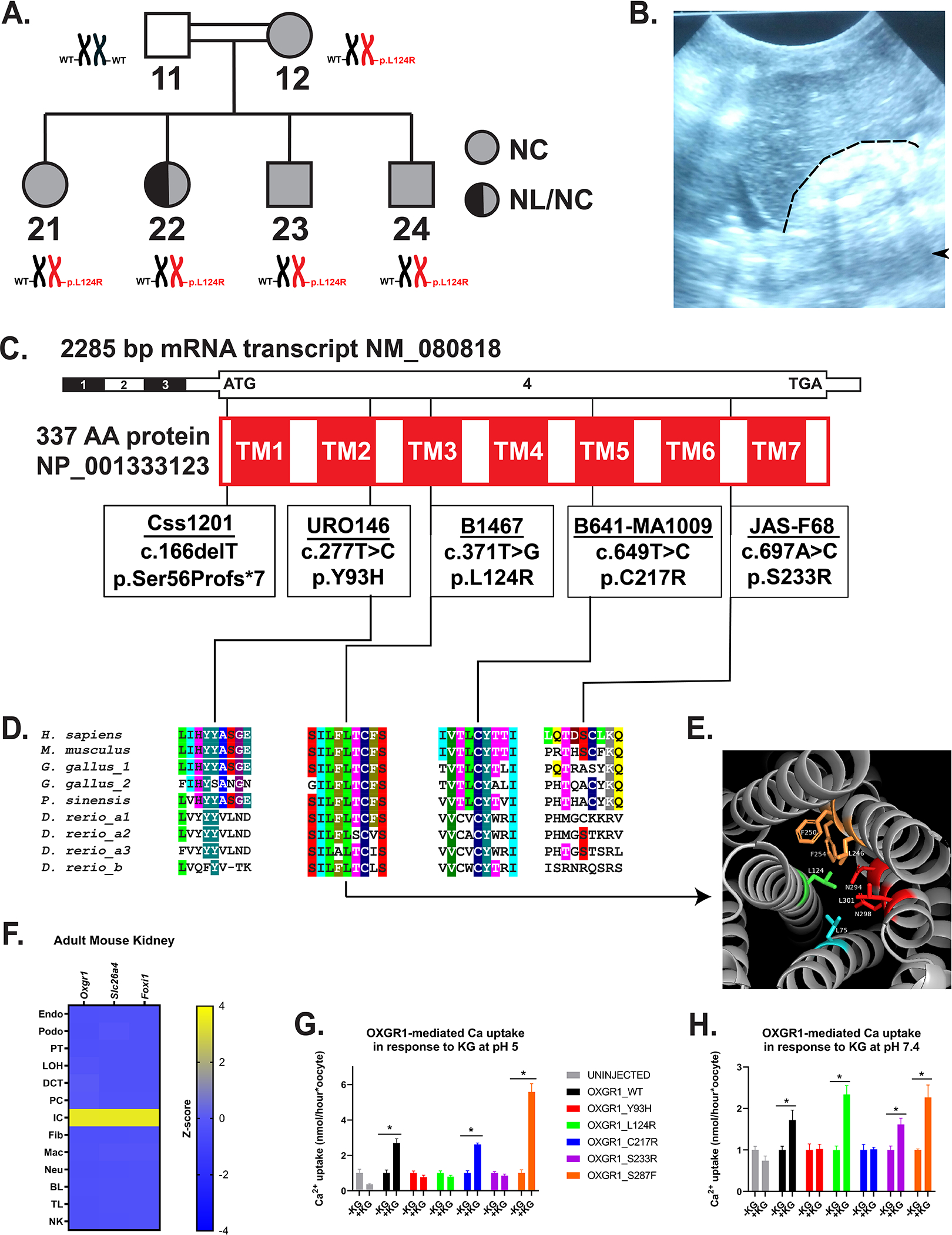
(A) Pedigree of Family B1467 with individual identifiers below symbols indicating absence or presence of NL and/or NC disease. Adjacent to each individual symbol is OXGR1 genotype.
(B) Renal ultrasound from subject B1467_21 shows increased echogenicity of right kidney (to right of and below dashed line) relative to adjacent liver with post-acoustic shadowing (arrowhead).
(C) Schematics of exon (black-white) and protein domain (red-white) structures of OXGR1. Black vertical lines indicate positions of variants (in boxes).
(D) Evolutionary conservation of amino acid sequence for each amino acid position impacted by OXGR1 missense variants. Protein sequences of putative orthologues from rodents (Mus musculus), birds (Gallus gallus), reptiles (Pelodiscus sinensis), and fish (Danio rerio) were used. Invertebrate orthologues have not been identified.
(E) Predicted human OXGR1 structure shown. Leucine 124 (stick model) present on the central transmembrane domain 3 (TM3, green) and protrudes into the core of the 7-transmembrane domain structure. It is within 4Å of seven amino acids, of which five are hydrophobic.
(F) Oxgr1 mRNA (z-score) was predominantly expressed in the intercalated cell (IC) cluster from single-cell mRNA sequencing data from adult mouse kidneys. Oxgr1 expression was highest in the IC cluster marked by expression of Slc26a4 and Foxi1, relative to other nephron tubular segment clusters to the left and interstitial or hematologic cell types to the right. Endo, endothelial cell; Podo, podocyte; PT, proximal tubular cell; LOH, loop of Henle cell; DCT, distal convoluted tubule; PC, principal cell; IC, intercalated cell; Fib, fibroblast; Mac, macrophage; Neu, neutrophil; BL, B lymphocyte; TL, T lymphocyte; NK, natural killer cell.
(G) Ca2+ uptake into Xenopus oocytes injected 48–72 hours previously with water or with cRNA (40 ng) encoding wild type human OXGR1 or the indicated variants. Uptake was measured at bath pH 5.0 for 30 min in the absence (−KG) or presence (+KG) of bath alpha-ketoglutarate (1 mM). Uptake measurements (nmol/hour) were normalized to the mean of the −KG group for each construct. *p<0.05 by Student’s t-test.
(H) Ca2+ uptake was measured as in (G) at bath pH 7.4. Uptake measurements were normalized to the mean of the −KG group for each construct. *p<0.05 by Student’s t-test.