
Fig. 7.
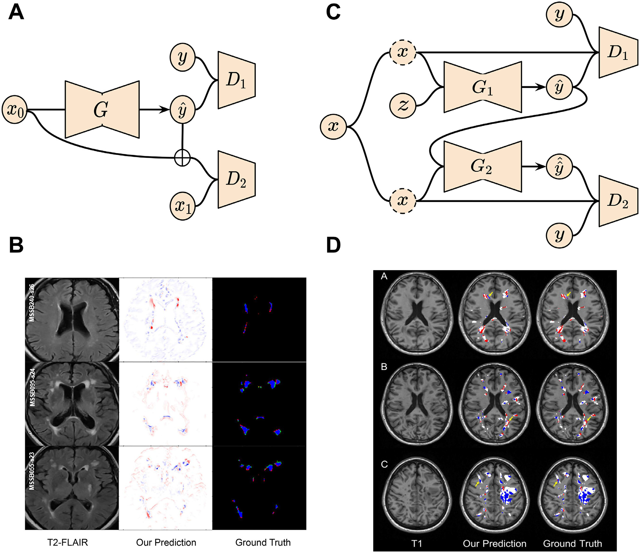
GAN applications in brain lesion (white matter hyperintensities and multiple sclerosis) evolution prediction. (A) Schematic of the GAN for white matter hyperintensities evolution prediction. (B) Disease evolution map examples produced by GAN and the derived irregularity map from two time points. (C) Two-stage conditional GAN for [11 C] PIB PET images generation from multi-sequence MR images for myelin content in multiple sclerosis dynamic prediction. (D) Examples of myelin content changes indicating demyelination (red color) and remyelination (blue color) from both GAN outputs and real PET images. Images are taken and adapted from Wei et al. (2020), Rachmadi et al. (2020).