
Fig. 8.
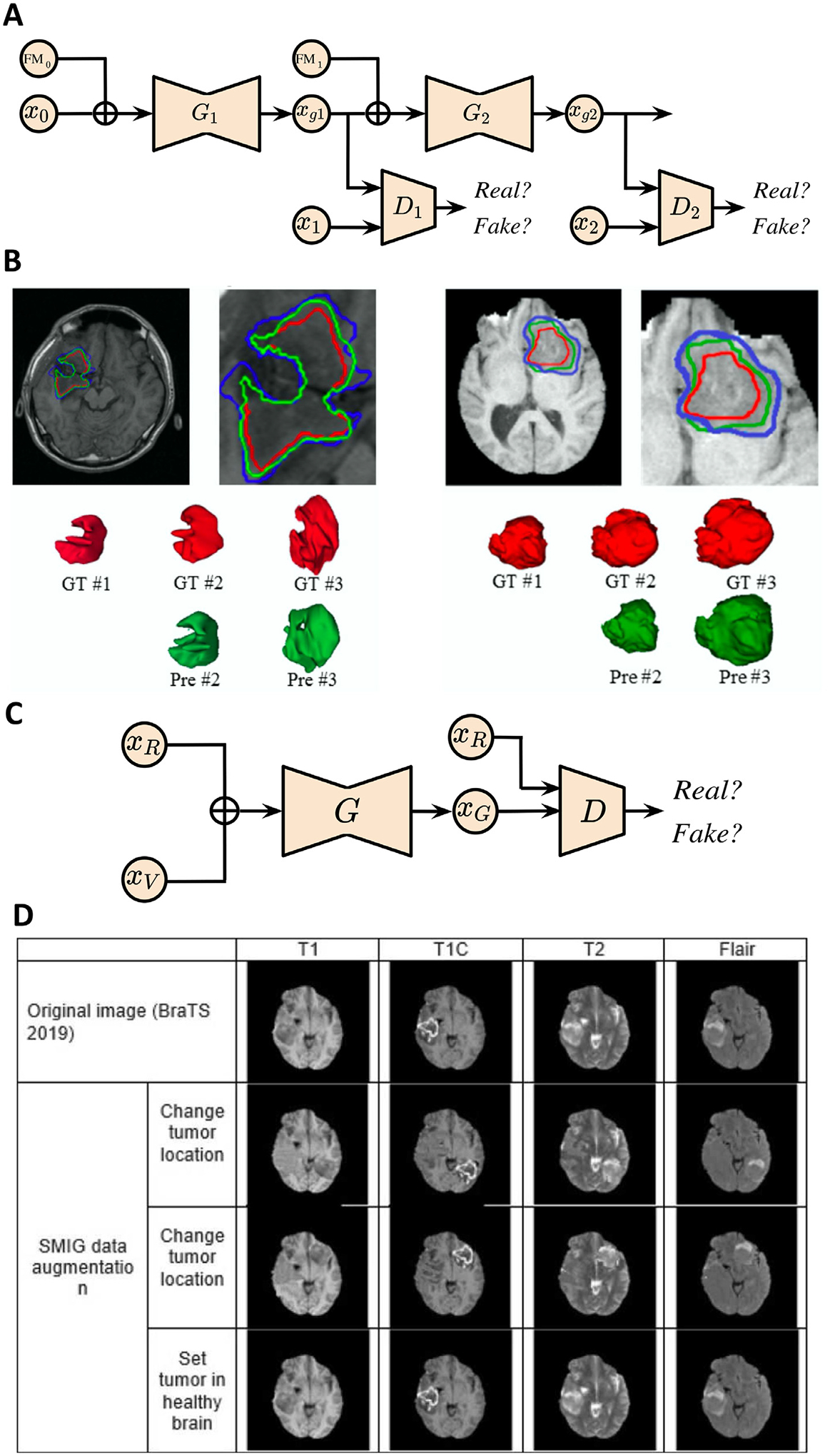
GAN applications in brain tumor growth prediction. (A) GP-GAN architecture for glioma growth prediction. xgi : generated image at time point i; Gi : generator at time point i; Di: discriminator at time point i. (B) Growth prediction for subjects with low-grade glioma (left) and high-grade glioma (right) at different time points via GP-GAN. GT: ground truth; Pre: prediction. (C) Schematic of SMIG model. The model is trained to 1) generate an abnormal brain based on a healthy brain from ADNI dataset and tumor volume from TCIA; 2) change tumor location. xR : image represents a healthy brain or tumor in real location; xV : tumor volume provided by TCIA; xG: generated image; G: generator; D: discriminator. (D) SMIG model applications on single patient images from BraTS dataset. Images are taken and adapted from Elazab et al. (2020), Kamli et al. (2020).