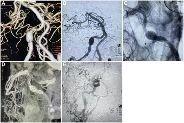
Figure 1.
Tubridge treatment on vertebral artery dissecting aneurysm and the follow-up. (A) CTA scan showing the dissecting aneurysm in the right vertebral artery, section 4 (V4); (B) DSA showing the dissecting aneurysm in the right vertebral artery, section 4 (V4); (C) Release of Tubridge flow diverter (3.5 × 55 mm); (D) DSA showing a goof wall apposition after final deployment of the flow diverter; (E) Follow-up 6 month after the treatment found no silhouette of the dissecting aneurysm.

