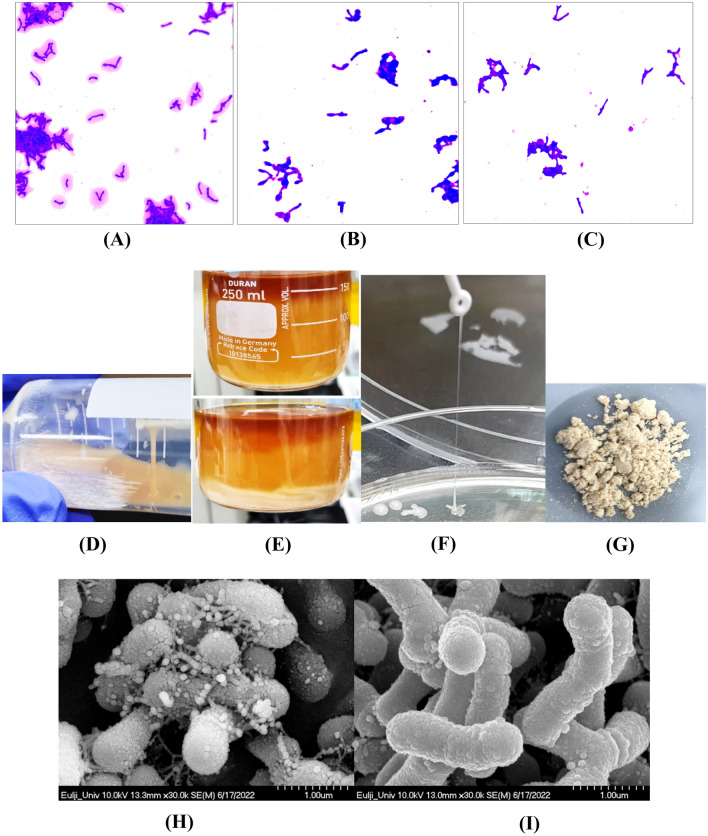
Fig. 1.
Visualization of EPS produced by B. bifidum EPS DA-LAIM. A Crystal violet staining of B. bifidum EPS DA-LAIM cultured in BL + 5% sucrose; B Crystal violet staining of B. bifidum EPS DA-LAIM cultured in BL + 5% glucose; C Crystal violet staining of B. bifidum DLP1224 cultured in BL + 5% sucrose; D Ropiness of B. bifidum EPS DA-LAIM culture broth; E three-layer separation of culture medium-EPS-bacteria cells in the culture broth F Ropiness of EPS from B. bifidum EPS DA-LAIM colony; G Freeze-dried EPS powder; H Scanning electron microscope’s image of the culture medium of B. bifidum EPS DA-LAIM; I Scanning electron microscope’s image of B. bifidum EPS DA-LAIM treated by centrifugation and washing with PBS (× 10,000)

