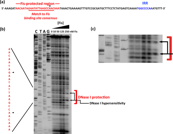
Fig. 2.
Identifying a binding site for Fis within fimS. (a) The DNA sequence of the right end of fimS in phase OFF, showing the location of the right inverted repeat, IRR (blue), and a sequence matching the consensus for Fis binding sites (red). (b) DNase I footprinting was performed with purified Fis protein and a DNA fragment corresponding to the right end of phase OFF fimS. Concentrations of Fis are given above each lane. The products of dideoxy chain-terminator nucleotide sequencing reactions, carried out with the same DNA fragment, are shown in the lanes labelled C, T, A and G. The assay revealed a region in which Fis protected the DNA from DNase I digestion, with two nucleotides exhibiting hypersensitivity to the enzyme. The protected region is highlighted with a red bracket and black arrows indicate the two hypersensitive bases. Black arrowheads point to the regions of hypersensitivity in the sequence shown on the left in red; this sequence corresponds to that shown in red in (a). This part of the image is reproduced in an enlarged format in (c).

