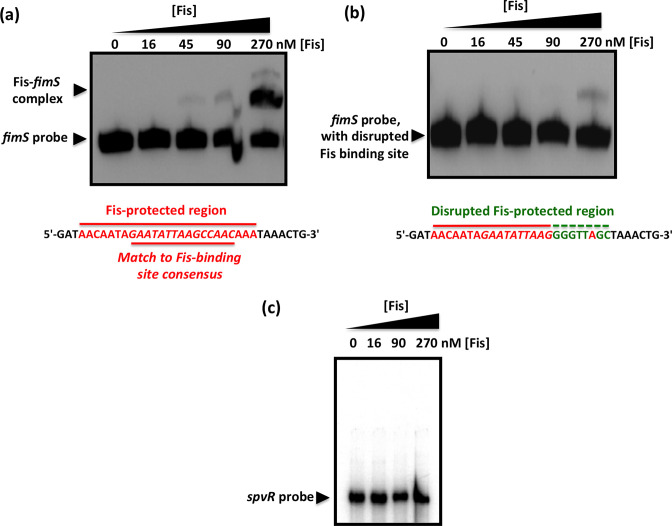
Fig. 3.
Mutation of the Fis binding site abrogates the Fis-mediated electrophoretic mobility shift of fimS DNA. (a) EMSA showing the gel mobility shift of a labelled 135 bp fragment of fimS DNA that includes the Fis binding site, highlighted in red below the gel. (b) Disruption of the Fis binding site by substituting the bases, shown in green below the gel, of the wild-type binding site sequence (red) almost completely abrogated the ability of Fis to alter the electrophoretic mobility of this DNA fragment. (c) A control EMSA using a 157 bp DNA fragment corresponding to the spvR promoter region from S. Typhimurium. This DNA fragment is known not to bind Fis [63]. Purified Fis failed to alter the electrophoretic mobility of the spvR DNA fragment at the same protein concentrations used in (a). The concentrations of purified Fis used in the experiments are given above each gel lane; black arrowheads indicate the bands corresponding to the unbound DNA probes and in (a), the Fis–fimS complex.

