Fig. 5.
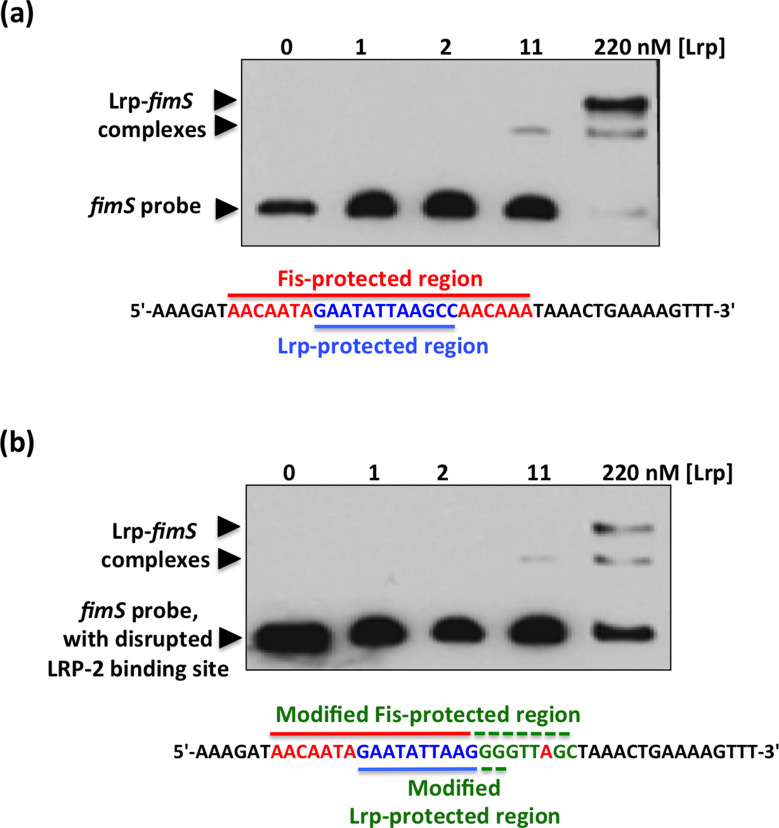
EMSA showing Lrp binding to fimS with an intact or a disrupted Fis binding site. (a) Purified Lrp protein was incubated at the concentrations shown with a PCR-generated 135 bp DNA fragment from fimS that contained an intact Fis binding site. The sequence of the region protected from DNase I digestion by Fis is shown in red and the Lrp binding site (LRP-2) is shown in blue. (b) The EMSA was repeated using the fimS derivative with the disrupted Fis binding site. The base substitutions are shown below the gel in green, together with the unchanged bases from the Fis binding site (red) and the LRP-2 site (blue). In both (a) and (b), arrowheads show the positions of bands corresponding to the unbound DNA probe and the Lrp–fimS complexes.

