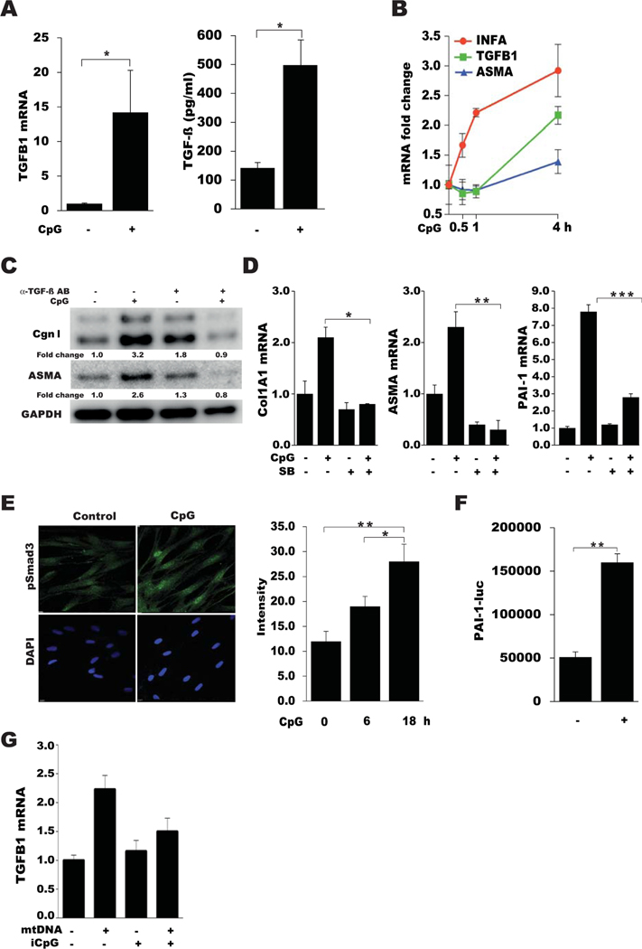
Figure 5.
TLR9-mediated fibrotic responses are transforming growth factor β (TGFβ) dependent. A–E, Confluent skin fibroblasts were incubated in media with CpG alone or with TGFβ. A, Left, Levels of TGFB1 mRNA as determined by qPCR. Values are the mean ± SEM of triplicate experiments. Right, Levels of TGFβ secreted in medium, as determined by enzyme-linked immunosorbent assay. B, Fold changes in mRNA levels at the indicated time points. Values were normalized to GAPDH mRNA values and are the mean ± SEM of triplicate determinations. C, Western blot analysis of whole cell lysates, treated with or without anti-TGFβ antibodies (AB). Representative immunoblots are shown. D, Expression of COL1A1, ASMA, and PAI1 mRNA in cultures preincubated with or without SB431542 (SB) for 60 minutes, as determined by qPCR. Values are the mean ± SEM of triplicate determinations. E, Left, Representative immunofluorescence microscopic images of confluent skin fibroblasts stained with anti-pSmad3 antibodies (green) and DAPI (blue), in the absence or presence of CpG. Original magnification × 100. Right, Quantification of nuclear pSmad3 phosphorylation at different time points. Values are the mean ± SD of 5 randomly selected high-power fields. F, Plasminogen activator inhibitor (PAI-1)–Luc activity in cell-free conditioned medium from fibroblasts incubated with or without CpG. Values are the mean ± SEM of triplicate determinations and are representative of 3 independent experiments. G, Expression of TGFB1 mRNA in fibroblasts incubated with mitochondrial DNA (mtDNA) in the absence or presence of iCpG. Values are the mean ± SEM. * = P < 0.05; ** = P < 0.01; *** = P < 0.001. See Figure 4 for other definitions.

