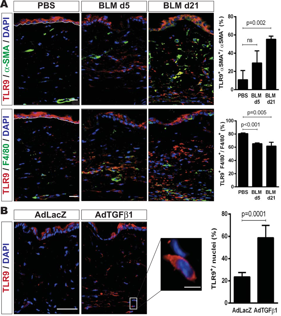
Figure 6.
Skin fibrosis in murine scleroderma is accompanied by up-regulated Toll-like receptor 9 (TLR9) expression. A, Left, Representative immunofluorescence microscopic images of lesional skin harvested on day 5 (d5) or day 21 from mice that received daily subcutaneous injections of bleomycin (BLM) or phosphate buffered saline (PBS), showing staining for TLR9 (red), α-smooth muscle actin (α -SMA) (green), or F4/80 (green). Nuclei were stained with DAPI (blue). Bars = 20 μm. Right, Quantification of cells double-positive for TLR9 and α -SMA (top) or TLR9 and F4/80 (bottom). Values are the mean ± SEM percentage. B, Left, Representative immunofluorescence microscopic images of lesional skin harvested on day 42 from mice that received a single injection of AdTGFβ1 or AdLacZ, showing staining for TLR9. Nuclei were stained with DAPI. Bar = 20 μm; bar in higher-magnification view of boxed area = 10 μm. Right, Quantification of the number of TLR9-positive cells per nuclei in 3 high-power fields per mouse. Values are the mean ± SEM percentage (≥3 mice per treatment group).

