Fig. 4.
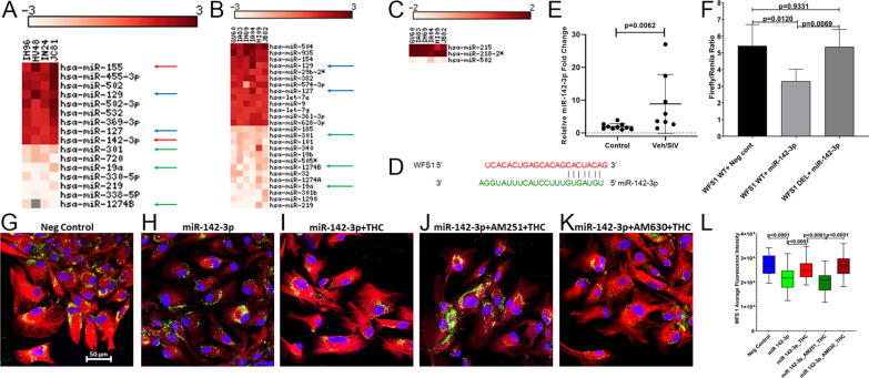
THC can counteract the post-transcriptional silencing of WFS1 by miR-142-3p. The heat map shows all differentially expressed (p ≤ 0.05) miRNAs in the BG of VEH/SIV (A) and THC/SIV (B) relative to uninfected controls RMs and in VEH/SIV compared to THC/SIV RMs (C). MiRNA species originating from the opposite arm of the precursor are denoted with an asterisk (*). Red arrows (A) indicate inflammation-associated miRNAs differentially upregulated in BG of VEH/SIV RMs. Blue and green arrows indicate miRNAs commonly up and downregulated in BG of VEH/SIV (A) and THC/SIV (B) RMs, respectively. MiRNA–mRNA duplex showing a single miR-142-3p binding site on the RM WFS1 (D) mRNA 3’ UTR. RT-qPCR validation of miR-142-3p expression in BG of VEH/SIV relative to uninfected control RMs (E). Luciferase reporter vectors containing a single highly conserved miR-142-3p (F) binding site on the RM WFS1 mRNA 3′ UTR or the corresponding construct with the binding sites deleted (WFS1 DEL) were co-transfected into HEK293 cells with 30 nM miR-142-3p or negative control mimic. Firefly and Renilla luciferase activities were detected using the Dual-Glo luciferase assay system 96 h after transfection. Luciferase reporter assays were performed thrice in six replicate wells (F). Representative immunofluorescence images showing the expression of WFS1 (red) protein at 96 h post-transfection of HCN2 neuronal cells with 30 nM LNA-conjugated FAM-labeled negative control (G) or miR-142-3p (green) (H) mimics and images were quantitated (L). miR-142-3p transfected HCN2 cells were treated with DMSO (H), THC (I), AM251 + THC (J), AM630 + THC (K) 96 h post-transfection. Cells were fixed and stained after 18 h and the expression of WFS1 (red) protein and nuclear staining using DAPI (blue) were quantitated (L). Experiments were performed in triplicate wells using 3 µM of THC, 10 µM of AM251/AM630, and repeated thrice. Firefly/Renilla ratios and immunofluorescence data were analyzed using one-way ANOVA followed by Tukey’s multiple comparison post hoc test. A p-value of < 0.05 was considered significant

