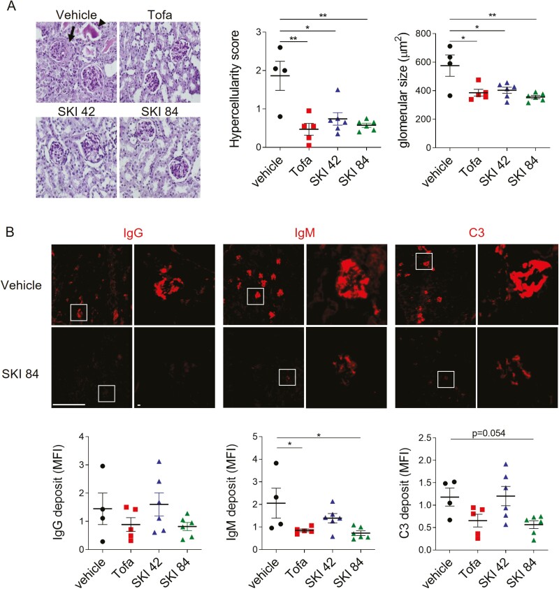
Figure 4:
histopathological alteration of kidney tissues by SKI-O-703 in NZB/W mice. Female NZB/W F1 mice were administrated orally with 42 mpk SKI-O-703 (SKI 42), 84 mpk SKI-O-703 (SKI 84), or tofacitinib (Tofa) from 18 to 34 weeks of age, and kidneys were examined at 34 weeks by histopathological methods. (A) Paraffin sections were stained with PAS and hematoxylin. An eosinophilic protein cast and crescent are indicated by the arrowhead and arrow, respectively. The images are representative of each group. Graphs show means ± SEMs with symbols representing values of individual mice. (B) Cryosections were stained with anti-IgG, anti-IgM, and anti-C3 Abs and observed by fluorescence confocal microscopy. Boxes in the left images are magnified in the right images. Representative images with mean fluorescence intensities are shown. Bar scale, 512 μm. *P < 0.05 and **P < 0.01 by two-tailed unpaired Students t-test.

