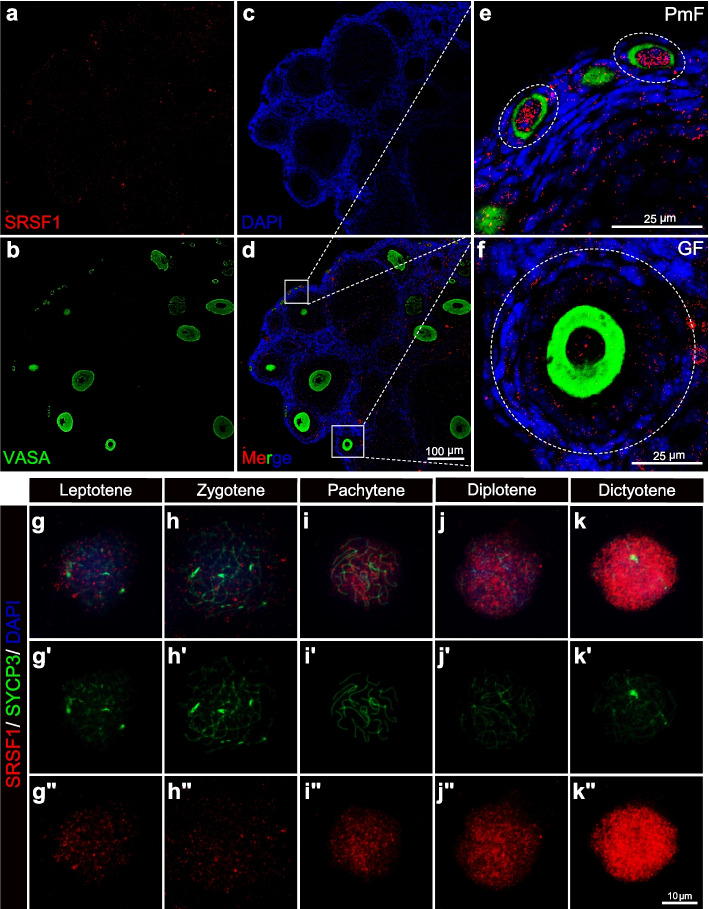
Fig. 1.
Dynamic localization of SRSF1 in mouse oocytes. a–f SRSF1 is highly expressed in oocytes of the primordial follicle. Immunostaining was performed using VASA and SRSF1 antibodies from adult mouse ovaries. DNA was stained with DAPI. a–d Scale bar, 100 μm. The e primordial follicle (PmF) and f grown follicle (GF) are shown in the magnified views. Scale bar, 25 μm. g–k The dynamic localization of SRSF1 to the oocyte meiotic prophase I program. Co-immunostaining was performed using SYCP3 and SRSF1 antibodies from 15.5 dpc, 17.5 dpc, 18.5 dpc, and 1 dpp oocyte surface spreading. DNA was stained with DAPI. gʹ–kʹ The meiotic stages of the oocytes were determined by SYCP3 staining (green). gʺ–kʺ The dynamic localization of SRSF1(red) during the oocyte meiotic prophase I program. Scale bar, 10 μm

