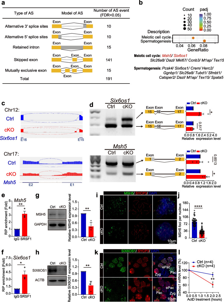
Fig. 7.
SRSF1 directly regulates the splicing of Msh5 and Six6os1. a Five AS events were significantly affected by SRSF1-deficient oocytes. rMATS (3.2.5) software was used to analyse AS events (FDR < 0.05). FDR, false discovery rate calculated from the P value. b Scatter plot of the GO enrichment analysis of the 191 affected AS events. c A schematic of the regulation of the splicing of Msh5 and Six6os1. Integrative Genomics Viewer (IGV, version 2.10.2) was used to visualize and confirm AS events in RNA-seq data. E, exon. d The ectopic splicing of Msh5 and Six6os1 in cKO ovaries was analysed by RT–PCR (n = 4 per group). The scheme and cumulative data on the percentage of the indicated fragment are shown accordingly. e, f SRSF1 directly regulated the expression of Msh5 and Six6os1 by RIP–qPCR in 16.5 dpc mouse ovaries. n = 3. g, h Western blotting of MSH5 and SIX6OS1 expression in 17.5 dpc Ctrl and cKO ovaries. GAPDH (g) or ACTB (h) served as a loading control. n = 4. i Localization of MSH5 and SYCP3 in 17.5 dpc Ctrl and cKO oocytes. DNA was stained with DAPI. Scale bar, 10 μm. j The number of MSH5 foci (green) was significantly reduced in cKO pachytene oocytes compared with Ctrl oocytes. Sixty Ctrl oocytes and sixty cKO oocytes were obtained from 4 animals. k Co-immunostaining of SIX6OS1 and SYCP3 in 17.5 dpc Ctrl and cKO ovaries. DNA was stained with DAPI. Zyg, zygotene; ePac, early pachytene; Pac, pachytene. Scale bar, 10 μm. l The expression of Six6os1 in 16.5 dpc cKO ovaries after ActD treatment at different times. n = 4. Significance was determined by unpaired Student’s t test; detailed P value P ≥ 0.05, *P < 0.05, ***P < 0.001, ****P < 0.0001. The error bar represents the mean ± SEM

