Fig. 5.
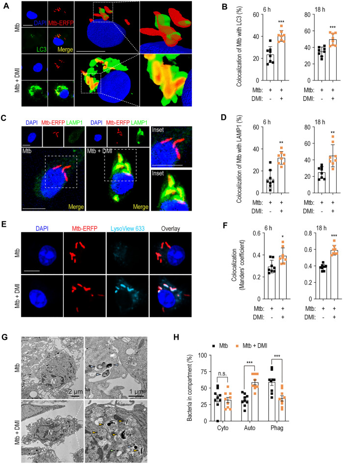
DMI enhances antibacterial autophagy against infection with Mtb in BMDMs. A–D BMDMs were infected with Mtb-ERFP (MOI 5) and followed by treatment with SC or DMI (100 μM) for the indicated times. Mtb-ERFP (red), Alexa Fluor 488-conjugated LC3 (green, A) or LAMP1 (green, C), DAPI (for nuclei, blue) were detected by confocal microscopy. Representative immunofluorescence images (A for LC3, C for LAMP1) and quantitation of colocalization of Mtb-ERFP with LC3 (B) or LAMP1 (D) were shown. Scale bars, 2 μm. E, F Mtb-ERFP-infected (MOI 5) BMDMs were treated with SC or DMI (100 μM) for the indicated times. Mtb-ERFP (red), Lysoview 633 (skyblue), and DAPI (for nuclei, blue) were detected by confocal microscopy. Representative immunofluorescence images (E) and quantitation of colocalization of Mtb-ERFP with LysoView 633 (F) using Manders’ coefficient were assessed. G, H BMDMs were infected with Mtb (MOI 5) and followed by treatment with SC or DMI (100 μM) for 18 h. Representative TEM images (G) and quantitation of bacteria in compartment (H). Bacteria in cytosol (light green), autophagosomes (orange), and phagosomes (pale blue) were marked as indicated. Unpaired Student’s t-test was used to examine the statistical analysis and the results were shown as means ± SD from at least three independent experiments performed. N nucleus, DMI dimethyl itaconate, n.s. not significant, Cyto cytosol, Auto autophagosomal/autolysosomal structure, Phag phagosome. *p < 0.05, **p < 0.01, and ***p < 0.001

