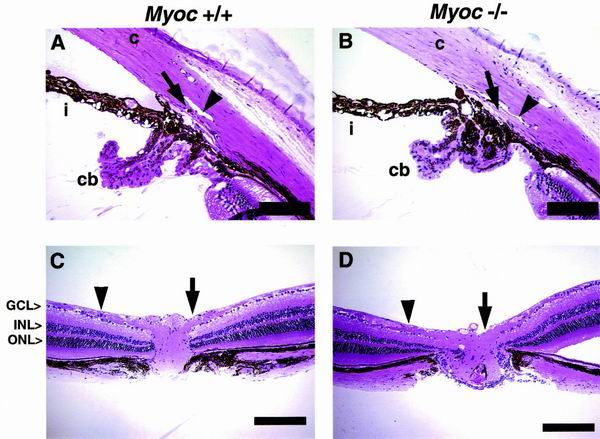
FIG. 5.
Analysis of iridocorneal angle, retina, and optic nerve. Histological examinations revealed no distinct abnormalities of eyes collected from all three genotypes of age-matched animals. (A and B) Anterior segment. The Schlemm's canal (arrowhead), trabecular meshwork (arrow), cornea (c), iris (i), and ciliary body (cb) are well developed and appear to be normal in −/− tissue. The Schlemm's canal extends from a region above the posterior ciliary body to a region near the posterior cornea. An iris process attaches to the anterior trabecular meshwork in many cases. (C and D) The retina including optic nerve fibers and optic nerve head. The RGC layer (GCL) is approximately one to two cells thick, and the thickness and number of cells within the inner nuclear layer (INL) and outer nuclear layer (ONL) appear unchanged in −/− tissue. Nerve fibers (arrowheads) run laterally and converge at the optic nerve head (arrows), through which they exit the retina. The thickness of nerve fibers and the size of optic nerve head also appear normal in −/− eyes. All the histological examinations performed on age- and background-matched +/− mice also revealed no discernible phenotypes (data not shown). Scale bars: 100 μm (A and B) and 200 μm (C and D).

