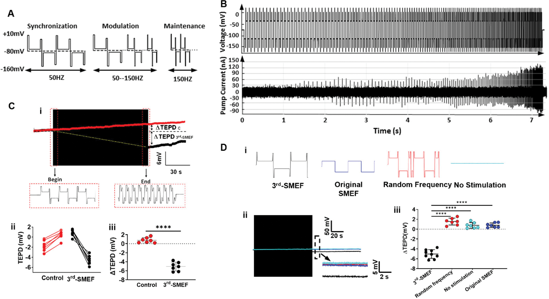
Figure 2. The 3rdgen-SMEF maintains the TEPD of ATP-depleted kidneys.

(A). Waveform of all three phases for the 3rdgen-SMEF applied: phase 1 synchronization (left), phase 2 modulation (middle), and phase 3 maintenance (right). (B). Pump currents resulting from this oscillating electrical field. The synchronization frequency was gradually increased in a stepwise pattern (change of 5% to 10% for every 10 to 20 oscillating pulses) up to the target frequency of 150 Hz. The increasing field frequency was accompanied by increases in the magnitude and density of the transient pump currents. (C). The TEPD of PCTs in the isolated Sprague Dawley rat kidneys in the presence (black trace in subpanel i) and absence (red trace in subpanel i) of 3rdgen-SMEF. TEPD was measured with a microelectrode, which was placed in the proximal tubule of the isolated kidneys. The black box was the compressed oscillating electric field of the 3rdgen-SMEF. Panels ii and iii summarized the results from the experiments performed. Significant differences were determined with student t-test. (****P<0.0001, n = 7 kidneys/group) (D). Effects of different electric fields (subpanel i, no stimulation-teal; random frequency-red; original SMEF-blue; and 3rdgen-SMEF-black) on TEPD in PCTs. Detailed TEPD changes right after the application of the different electric fields (black box) were measured in the dash-line box shown as the inserts in D-ii. The results were summarized in D-iii. Results are presented as mean ± SD. Significant differences were determined with one-way ANOVA followed by Dunnett’s multiple comparisons test. (****P<0.0001, n = 7–9 kidneys/group).
