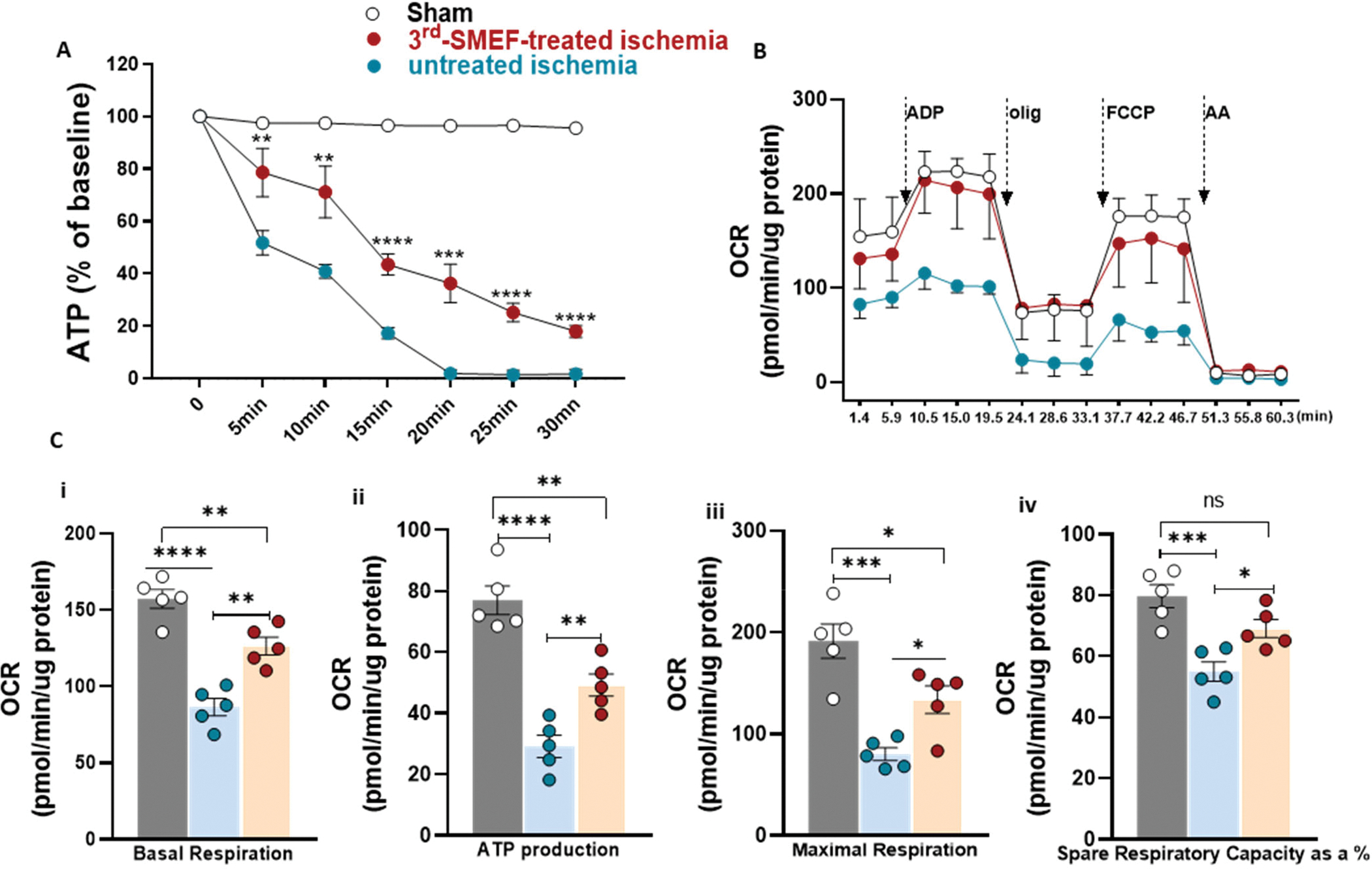
Figure 3. ATP content and mitochondrial function are preserved with the application of accelerating-3rdgen-SMEF.

(A). ATP content in kidney tissue was measured at different time points of ischemia without reperfusion. Both renal pedicles were clamped, one as untreated control and the other one treated with accelerating-3rdgen-SMEF. Data are presented as mean ± SEM. Two-way ANOVA followed by Tukey multiple comparisons test have been performed (**p<0.01; ***p < 0.001; ****p<0.0001, n = 5 mice/group). (B) Oxygen consumption rate (OCR) traces of isolated mitochondria from C57BL/6J mouse kidneys underwent 15 min of ischemia with or without accelerating-3rdgen-SMEF treatment, expressed as picomoles of O2 per minute, under basal conditions and after the injection of ADP (1mM), oligomycin (2 μM), FCCP (4 μM), and AA+ rotenone (2 μM). Oligo, oligomycin; FCCP, carbonyl cyanide 4-[trifluoromethoxy]phenylhydrazone; AA, antimycin A. (C). Analysis of mitochondrial respiratory parameters obtained from normalized XFe24 graphs (B). Data are presented as mean ± sem. One-way ANOVA followed by Tukey multiple comparisons test have been performed (*p<0.05; **p<0.01; ***p < 0.001; ****p<0.0001, n = 5 kidneys/group).
