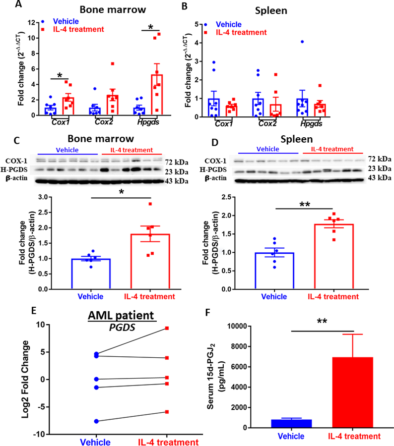
Figure 2. IL-4 supplement activates prostaglandin metabolism.

(A)(B) Expression of genes (Cox1, Cox2, and Hpgds) in the prostanoid biosynthesis pathway assessed by qPCR analysis in the bone marrow (A) and spleen (B) of AML mice. Data were normalized to 18S rRNA expression. (n=6~8). Western blot showing expression of COX-1 and H-PGDS in cells isolated from bone marrow (C) and spleen (D) of AML mice. Densitometry was done by normalizing to vehicle control and relative to β-actin for H-PGDS. Data shown are mean ± SEM per group, each group has at least three replicates. (n=6). (E) Expression of PGDS in patient-derived AML cells treated with vehicle or IL-4 (n=5). (F) Serum level of 15d-PGJ2 of AML mice treated with vehicle (n=10) or IL-4 (n=8) measured by ELISA. Data represent mean± SEM in A-D and F, unpaired two-tailed Student t test was utilized; and paired two-tailed Student t test was applied to E; *, P < 0.05; **, P < 0.01.
