
FIG. 6.
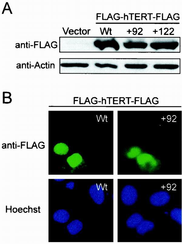
Protein stability and nuclear localization of hTERT with mutations in the DAT domain. (A) Anti-FLAG Western blot of lysates from 293T cells transiently transfected with biologically essential hTERT mutants +92, +122, wild-type hTERT, or control vector. The anti-actin blot shows equal protein loading. (B) Subcellular localization of DAT domain mutants transiently expressed in U2OS cells by indirect immunofluorescence. Localization of FLAG-hTERT-FLAG was visualized with an anti-FLAG antibody recognized by a fluorescein isothiocyanate-conjugated secondary antibody (green). Hoechst was used to stain nuclei (blue).