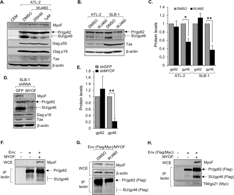
Fig 6. MyoF regulates the intracellular abundance of HTLV-1 Env.
(A) Inhibition of MyoF reduces levels of SU (gp46). ATL-2 cells were treated with DMSO or WJ460 (200 nM or 1 μM) for 24h. Whole cell extracts (50 μg) from uninfected CEM and the treated cells were analyzed by Western blot using antibodies against MyoF, β-actin and the viral proteins Tax, Gag p55, Gag p19, and SU/Pr. (B) ATL-2 and SLB-1 cells were treated with DMSO or 1 μM WJ460 for 24h. Whole cell extracts (100 μg for Tax; 50 μg for the others) were analyzed by Western blot using antibodies against β-actin and the viral proteins Tax, and SU/Pr. (C) The graph shows quantification of band intensities of Pr (gp62) and SU (gp46) normalized to band intensities of β-actin averaged from three and four independent experiments for ATL-2 and SLB-1 cells, respectively. Error bars show standard deviations; * p<0.05, ** p<0.01. (D) Knockdown of MyoF expression reduces levels of SU (gp46). Whole cell extracts were prepared from SLB-1 cells stably expressing an shRNA targeting GFP (negative control) or MYOF mRNA, and 50 μg from each cell line was analyzed by Western blot using antibodies against MyoF, β-actin and the viral proteins Tax, Gag p19, and SU/Pr. (E) The graph shows quantification of band intensities of Pr (gp62) and SU (gp46) normalized to band intensities of β-actin averaged from three independent experiments. Error bars show standard deviations; ** p<0.01. (F) Ectopic co-expression of MyoF with Env in HEK293T cells increases SU (gp46) abundance. Cells (1 x 10⁶) were transfected with pcDNA-GFP-HA-MyoF (3 μg) and pCMV-HTLV-1-Env (1 μg). Env was co-precipitated with Lens Culinaris Agglutinin lectin bound to agarose beads (P-lectin) from 879 μg of whole cell extracts (WCE) and analyzed along with 50 μg WCE by Western blot using antibodies against MyoF, and SU/Pr. (G) Inhibition of MyoF ectopically co-expressed with Env in HEK293T cells decreases SU/gp46 abundance. Cells (1 x 10⁶) were transfected with pcDNA-GFP-HA-MyoF (3 μg) and pCMV-HTLV-1-Env-Flag-Myc (3 μg) expression vectors and treated with DMSO or WJ460 (1 μM) for 24h. Env was co-precipitated with Lens Culinaris Agglutinin lectin bound to agarose beads (P-lectin) from 768 μg of whole cell extracts (WCE) and analyzed along with 50 μg WCE by Western blot using antibodies against MyoF, β-actin and the Flag-epitope tag of Env. (H) Ectopic co-expression of MyoF with Env in HEK293T cells increases TM (gp21) abundance. Cells (1 x 10⁶) were transfected with pcDNA-GFP-HA-MyoF (3 μg) and pCMV-HTLV-1-Env-Flag-Myc (3 μg). Env was co-precipitated with Lens Culinaris Agglutinin lectin bound to agarose beads (P-lectin) from 1190 μg of whole cell extracts (WCE) and analyzed along with 50 μg WCE by Western blot using antibodies against MyoF, and the Flag- and Myc-epitope tags of Env.

