SUMMARY
In our case series of monkeypox (MPX) virus infected patients, one had a single genital ulcer as the only cutaneous manifestation of the infection. Physical examination revealed a single, rounded ulcer of the shaft penis characterized by pinkish raised, infiltrated borders and a crusty yellowish bottom associated with bilateral inguinal lymphadenopathies. Serology for Treponema pallidum infection and a complete screening for sexually transmitted infections (STIs) resulted negative except for the detection of Staphylococcus aureus at the cultural examination and MPX DNA at the ulcer bottom. The patient’s general conditions were good therefore he remained isolated at home for 3 weeks after the diagnosis. At one month follow up, he presented only a depressed pinkish skin scars on the site of the previous ulcer. The clinical presentation of this patient could easily be misdiagnosed with other sexually transmitted infections (STIs), especially with primary syphilis. MPX infection should be considered in the differential diagnosis of STIs, also in patients with weak and localized manifestations.
Keywords: Monkeypox virus, sexually transmitted infections, genital ulcer
INTRODUCTION
Since May 2022, multiple human monkeypox virus (MPX) cases have been identified in countries outside the MPX endemic regions (Democratic Republic of the Congo and other countries of Central and West Africa). This new epidemic has surged mainly among men who have sex with men (MSM) and rapidly reaches the proportions of a global health emergency during the summer 2022 [1–3].
In our case series of 16 MPX infected patients diagnosed from 1th July until 31th August 2022 in the Dermatology and Infectious Disease Units of the San Martino Hospital, Genoa, one patient had a single genital ulcer as the only cutaneous manifestation of the infection.
CASE REPORT
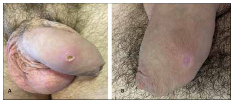
The patient was a 33-year-old Italian homosexual man presented at the sexually transmitted infections (STIs) outpatient clinic of the Dermatology Unit complaining of a single, asymptomatic, genital lesion appeared 10 days before, preceded by sore throat and neck swelling. His human immunodeficiency virus (HIV) status was negative and he used, on demand, the pre-exposure prophylaxis (PrEP) for HIV prevention. The patient had a history of secondary syphilis diagnosed and treated two years before with an enhanced treatment regimen consisting of the addition of doxycycline and ceftriaxone to the conventional benzathine penicillin G treatment [4]. Moreover, he had a history of anal high-risk human papillomavirus (HR-HPV) infection associated with normal cytology, diagnosed two months earlier with the HR-HPV testing, complementary to liquid-base anal cytology (Thin Prep®), as described elsewhere [5]. Physical examination revealed a single, rounded ulcer of the shaft penis characterized by pinkish raised, infiltrated borders and a crusty yellowish bottom (Figure 1A), associated with bilateral inguinal lymphadenopathies; oropharyngeal involvement was not observed but cervical lymphadenopathies, which made swallowing difficult, were present on palpation. The patient had travelled to Spain in the preceding three weeks, where he had unprotected sexual intercourses with casual partners. Suspecting first primary syphilis, we performed serology for Treponema pallidum infection and a complete STIs screening: serology for HIV, hepatitis B and C viruses, lesion and urethral swabs for DNA search of Chlamydia trachomatis, Neisseria gonorrhoeae, Mycoplasma hominis, Mycoplasma genitalium by polymerase chain reaction (PCR); lesion swab for the search of common germs-fungi (microscopic and cultural examination) and, lastly, a swab on the bottom ulcer and on the oropharynx for the search of MPX DNA by PCR. The laboratory tests resulted negative, except for the detection of Staphylococcus aureus at the cultural examination and MPX DNA at the ulcer bottom. Since the patient’s general conditions were good, he remained isolated at home for 3 weeks after the diagnosis. At one month follow up, he was completely healed and presented only a depressed pinkish skin scars on the site of the previous ulcer, that was unchanged at the 3-months follow up visit (Figure 1B).
Figure 1.
A) Single, rounded ulcer of the shaft penis characterized by pinkish raised, infiltrated borders and a crusty yellowish bottom; B) depressed pinkish skin scars in the site of the previous ulcer (3 month follow up visit).
DISCUSSION
The clinical presentation of our patient could easily be misdiagnosed with other STIs, especially with primary syphilis with bacterial superinfection (which could be responsible for the yellowish crust) or with pseudo-chancre [6]. Indeed, we suggest considering MPX infection in all at-risk patients presenting with traditional STIs signs/symptoms to avoid misdiagnosis.
Differently from the patients described by Quattri et al. who had single lesions without systemic involvement, our patient had also signs/symptoms of systemic involvement. Indeed, MPX virus traditionally causes a systemic infection: once acquired through close contact with skin/mucosal lesions, after replication at the inoculation site, it spreads to the local lymph nodes and bloodstream [7]. This incubation period lasts 7–14 days. Prodromal signs/symptoms usually occur 1–2 days before the appearance of skin/mucosal lesions [1, 8].
The presentation of MPX infection with a single skin lesion accounts for about 10% of all MPX cases [1]. We can speculate that in such patients the cutaneous/mucosal viral load was so low to cause only localized, single lesions. Regrettably, in our patient, we cannot quantitatively assess the MPX viral load in the clinical samples (lesion swab and blood) to confirm this hypothesis.
In conclusion, MPX infection should be considered in the differential diagnosis of STIs, also in patients with weak and localized manifestations.
Footnotes
Conflict of interest
None to declare.
Funding sources
None to declare.
REFERENCES
- 1. Thornhill JP, Barkati S, Walmsley S, et al. Monkeypox virus infection in humans across 16 countries - April–June 2022. N Engl J Med. 2022;387(8):679–691. doi: 10.1056/NEJMoa2207323. [DOI] [PubMed] [Google Scholar]
- 2. Farahat RA, Sah R, El-Sakka AA, et al. Human monkeypox disease (MPX) Infez Med. 2022;30(3):372–391. doi: 10.53854/liim-3003-6. [DOI] [PMC free article] [PubMed] [Google Scholar]
- 3. Amer FA, Hammad NM, Wegdan AA, et al. Growing shreds of evidence for monkeypox to be a sexually transmitted infection. Infez Med. 2022;30(3):323–327. doi: 10.53854/liim-3003-1. [DOI] [PMC free article] [PubMed] [Google Scholar]
- 4. Drago F, Ciccarese G, Broccolo F, et al. A new enhanced antibiotic treatment for early and late syphilis. J Glob Antimicrob Resist. 2016;5:64–66. doi: 10.1016/j.jgar.2015.12.006. [DOI] [PubMed] [Google Scholar]
- 5. Ciccarese G, Herzum A, Rebora A, Drago F. Prevalence of genital, oral, and anal HPV infection among STI patients in Italy. J Med Virol. 2017;89(6):1121–1124. doi: 10.1002/jmv.24746. [DOI] [PubMed] [Google Scholar]
- 6. Drago F, Ciccarese G, Molle MF, Parodi A. Ulcer of the penis due to mycoplasma hominis infection: an example of pseudo-chancre. Ital J Dermatol Venerol. 2022;157(1):113–114. doi: 10.23736/S2784-8671.21.06937-1. [DOI] [PubMed] [Google Scholar]
- 7. Quattri E, Avallone G, Maronese CA, et al. Unilesional monkeypox: A report of two cases from Italy. Travel Med Infect Dis. 2022;49:102424. doi: 10.1016/j.tmaid.2022.102424. [DOI] [PMC free article] [PubMed] [Google Scholar]
- 8. Wong L, Gonzales-Zamora JA, Beauchamps L, Henry Z, Lichtenberger P. Clinical presentation of Monkeypox among people living with HIV in South Florida: a case series. Infez Med. 2022;3(4):610–618. doi: 10.53854/liim-3004-17. [DOI] [PMC free article] [PubMed] [Google Scholar]


