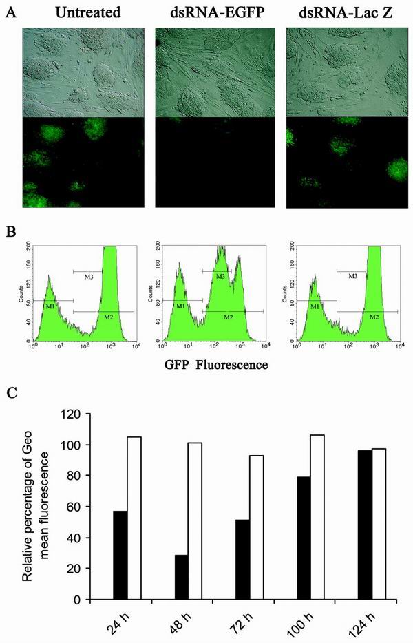
FIG. 7.
Sequence-specific inhibition of EGFP expression by dsRNA in the stable ES-EGFP clone, in which the EGFP gene is integrated as a single copy. (A) Fluorescence microscopy of undifferentiated ES clone untreated or transfected with 3 μg of in vitro-transcribed dsRNA-EGFP or dsRNA-lacZ, respectively. Photographs were taken 72 h later, using a bright field (upper panel) and fluorescence (lower panel). (B) FACS analysis of the ES clone 48 h after transfection. M1 indicates the gating of GFP-negative cells, M2 the gating of GFP-positive cells, and M3 the gating of cells with reduced fluorescence due to RNAi. Untransfected clone (left panel; geometric mean fluorescence: M1 = 6.12, M2= 743.15), cells transfected with dsRNA-EGFP (middle panel; geometric mean fluorescence: M1= 6.16, M2 = 270.93), cells transfected with dsRNA-lacZ (right panel; geometric mean fluorescence: M1 = 6.43, M2= 772.95). (C) Kinetics of RNAi in undifferentiated ES-EGFP clone. The relative geometric mean fluorescence of cells transfected with dsRNA-EGFP (solid bars) or dsRNA-lacZ (open bars) was normalized to the geometric mean fluorescence of untransfected cells. ES cells were split at 72 h after the initial transfection of dsRNA and plated at 2 × 10⁵ cells/well for the later time measurements at 100 and 124 h.

