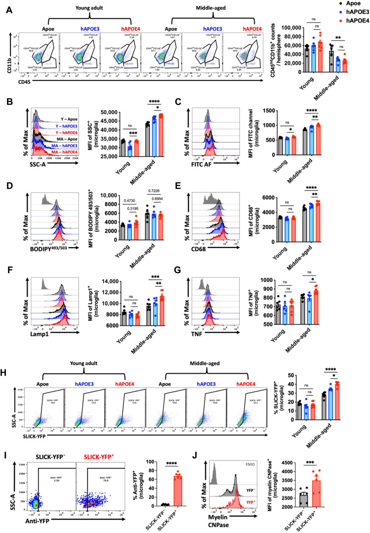
Fig. 2. APOE4 genotype promotes the emergence of the AF phenotype at middle age.
(A) Representative dot plots show the immune profile in the brains of young and middle-aged Apoe, hAPOE3, and hAPOE4, quantified on the right. Representative histograms depict the relative level of cell granularity (B), autofluorescence (C), lipid accumulation (D), CD68 (E), and Lamp1 (F) protein expression and intracellular cytokine production of TNF (G) in CD45intCD11b+ microglia across ages and genotypes. (H) Ex vivo neuronal engulfment assay shows a significant increase in the percent of microglia that phagocytized live SLICK-YFP neurons, quantified on the right. Validation of internalized myelinated cortical neurons was performed using intracellular detection of anti-YFP (I) and anti-myelin CNPase (J) in phagocytic (SLICK-YFP+) and nonphagocytic (SLICK-YFP−) microglial populations within the same brain. N = 6 to 7 per group. MA, middle-aged; Y, young; ns, not significant.. Data were analyzed using one-way analysis of variance (ANOVA) with Bonferroni post hoc correction for multiple comparisons (*P < 0.05, **P < 0.01, ***P < 0.001, and ****P < 0.0001).

