. 2023 Jan 27;12:e83602. doi: 10.7554/eLife.83602

Figure 6. The computed free energy of binding for blebbistatin from MSM-docking accurately predicts binding free energies for existing experimental data and for a myosin isoform whose blebbistatin sensitivity was not known.

(A) Predictions from MSM-docking are highly correlated to experimental values (R2=0.82) and most predictions are within 1 kcal/mol of experimental values. Error bars for predicted free energies of binding represent bootstrapped estimate of standard error of the mean from 250 trials. Error bars for experimental values show the standard error of the IC50 or Ki converted to a binding free energy. (B) An NADH-linked ATPase assay indicates that MYH7b is highly sensitive to blebbistatin inhibition (IC50: 0.36 μM), consistent with the prediction from MSM-docking. Data show the mean ATPase activity ± standard deviation across 5 experimental replicates (2 biological replicates, each with two or three technical replicates).

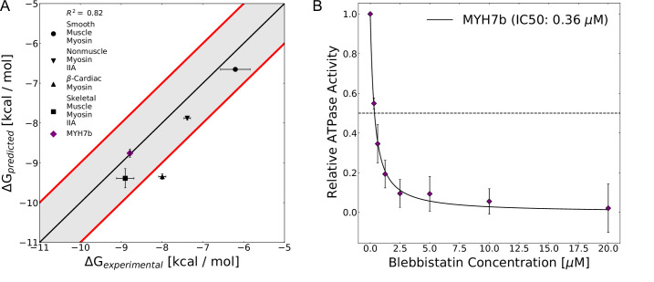
Figure 6.

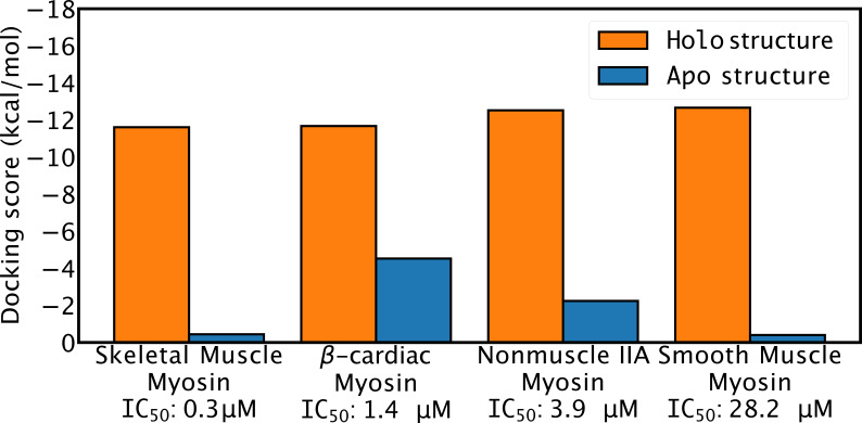
Figure 6—figure supplement 1. Docking scores to homology models of apo and holo structures do not correlate with blebbistatin potency.

Figure 6—figure supplement 1.

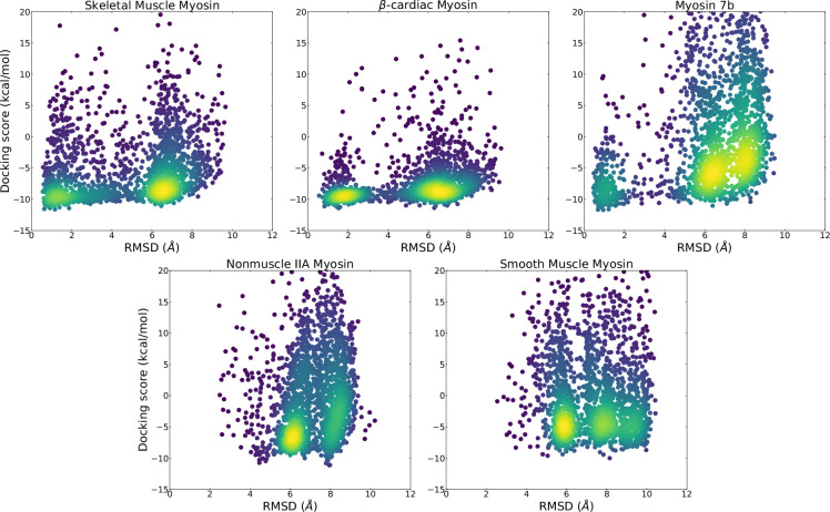
Figure 6—figure supplement 2. Comparison of distribution of docking scores and ligand heavy atom root mean square deviation (RMSD) from blebbistatin’s pose in a holo crystal structure (PDB: 1YV3) show that skeletal and β-cardiac myosin are more likely to adopt structures where blebbistatin can be docked in its holo orientation and obtain a favorable docking score.

Figure 6—figure supplement 2.

Points are colored by density with bright colors indicate areas of high density.
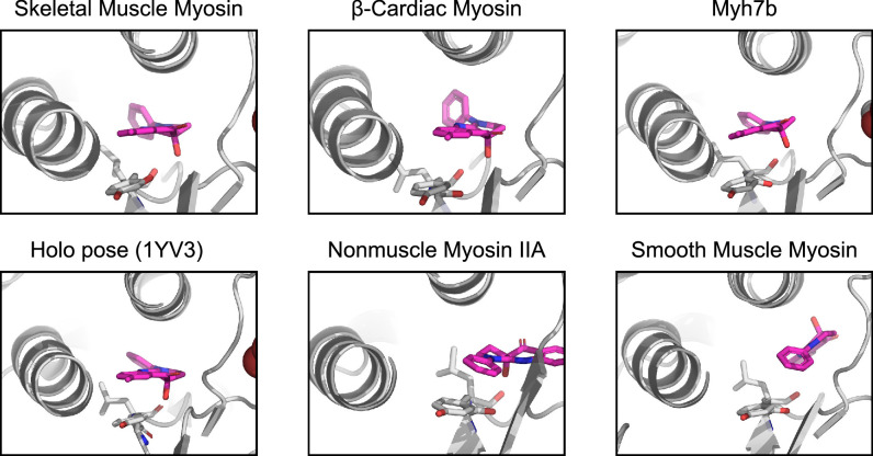
Figure 6—figure supplement 3. Highest scoring poses for each of the myosin isoforms reveals that the best pose for skeletal muscle myosin, β-cardiac myosin, and Myh7b closely matches the pose seen in holo crystal structures (ligand heavy atom RMSD 1.2 Å, 1.5 Å, and 0.9 Å to holo PDB 1YV3 for skeletal muscle myosin, β-cardiac myosin, and Myh7b, respectively).

Figure 6—figure supplement 3.

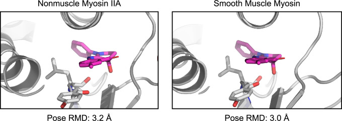
Figure 6—figure supplement 4. Docking to nonmuscle myosin IIA and smooth muscle myosin produces low RMSD poses (3.2 Å and 3.0 Å blebbistatin heavy atom RMSD from holo 1YV3 structure) with reasonably high scores (–6.3 kcal/mol in both cases).

Figure 6—figure supplement 4.

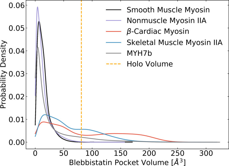
Figure 6—figure supplement 5. MSM-weighted pocket volumes for myosin-II isoforms in the ADP*Pi state reveal that blebbistatin pocket opening commonly occurs in skeletal muscle myosin, β-cardiac myosin, and Myh7b.

Figure 6—figure supplement 5.

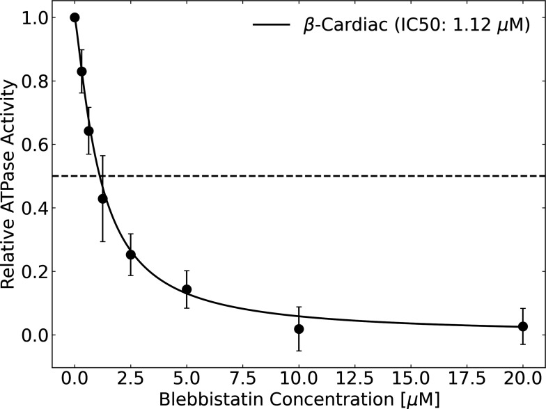
Figure 6—figure supplement 6. Blebbistatin inhibits the actin-activated ATPase activity of β-cardiac myosin with an IC50 of 1.12 μM.

Figure 6—figure supplement 6.

ATPase was measured using an NADH-linked assay. Error bars represent standard deviation across four trials (two biological replicates with two technical replicates each).