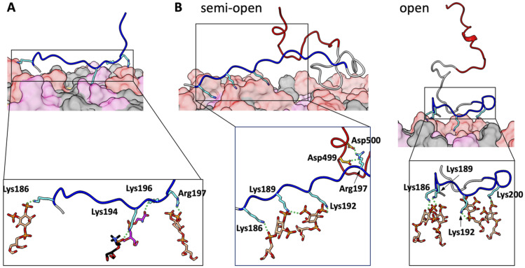
FIG. 3.
Representative snapshots from MD simulations of membrane-bound BR-only and BR-A fragments. (a) BR-only fragment fully bound to a membrane with 10% PIP2. (b) Two poses of the BR-A fragment bound to a membrane with 30% PIP2. In the semi-open pose, the first half of the BR is engaged with the membrane, whereas the second half of the BR interacts with the A motif. In the open pose, the A motif is fully released from the BR. Sidechains that form membrane contacts are shown as sticks. In the zoomed images, the lipid partners are also shown as sticks, as are sidechains that form salt bridges between the BR and A motif. The coloring scheme is the same as in Fig. 1; carbon atoms in a stick representation follow the same coloring scheme, except that those in the BR and A motif are in cyan and orange, respectively; O, N, and P atoms are in red, blue, and orange, respectively.

