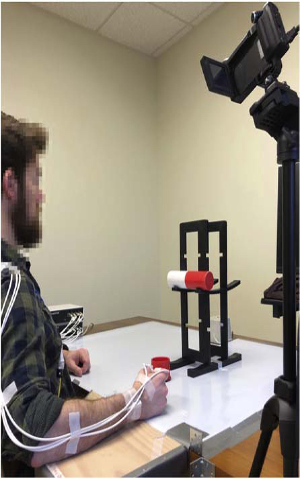
Figure 1. Participant and motor task setup.

A dowel rested horizontally on a cradle of a stand secured to the table. The dowel, positioned at 75% of the participant’s maximum arm reach, was centered to align with the participant’s acromion process. A target hole was centered in front of the apparatus at about 50% of the participant’s maximum reach. A taped line, positioned at 25% of the participant’s maximum reach, designated the starting position of the tested hand. Three electromagnetic markers were secured to the radial styloid and dorsal surface of the distal phalanges of the thumb and index fingers of the tested hand. An opaque screen (not pictured) occluded the participant’s view of the dowel. The video camera framed the tested hand and side view of the eyeline.
