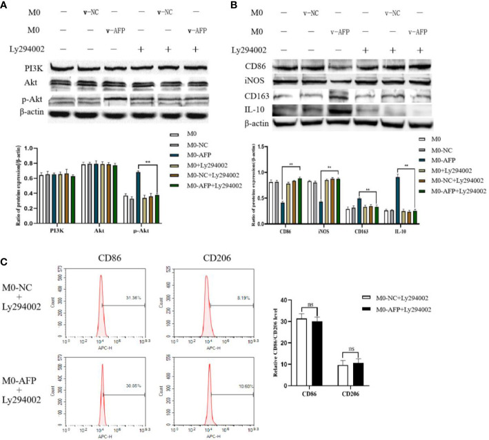
Figure 3.
The effects of AFP and the PI3K inhibitor Ly294002 on the expression of M1- and M2-type macrophages’ markers. The M0 macrophages were infected with AFP-expressing negative control lentivirus vectors (M0-NC) or AFP-expressing lentivirus vectors (M0-AFP) for 48 h and then treated with LPS+IFN-γ or IL-4+IL-13 for 24 h, followed by treated with Ly294002 (final concentration: 20 μmol/L) for 24 h. (A) The expression of PI3K, Akt and p-Akt(Ser473) proteins in the M0-NC and M0-AFP groups was analyzed by Western blotting; the right column diagram displays the expression levels of proteins in each group statistically analyzed by gray scanning, **P<0.01; (B) The expression of the M1-type macrophage markers CD86 and iNOS and the M2-type macrophage markers CD163 and IL-10 in the M0-NC and M0-AFP groups analyzed by Western blotting; the right column diagram displays the expression levels of proteins in each group statistically analyzed by gray scanning, **P<0.01. (C) The expression of the M1-type macrophage marker CD86 and the M2-type macrophage marker CD206 in macrophages of the M0-NC and M0-AFP groups was detected by flow cytometry; the right column diagram displays statistical analysis of the expression of CD86 and CD206 in each group (NS: P >0.05). The results of the at least three independent experiments are shown. v-NC: negative control lentivirus vectors; v-AFP: AFP-expressing lentivirus vectors.

